Wenn Sie das Fenster schließen, wird Ihre Konfiguration nicht gespeichert, es sei denn, Sie haben Ihren Artikel in die Bestellung aufgenommen oder zu Ihren Favoriten hinzugefügt.
Klicken Sie auf OK, um das MILLIPLEX® MAP-Tool zu schließen oder auf Abbrechen, um zu Ihrer Auswahl zurückzukehren.
Wählen Sie konfigurierbare Panels & Premixed-Kits - ODER - Kits für die zelluläre Signaltransduktion & MAPmates™
Konfigurieren Sie Ihre MILLIPLEX® MAP-Kits und lassen sich den Preis anzeigen.
Konfigurierbare Panels & Premixed-Kits
Unser breites Angebot enthält Multiplex-Panels, für die Sie die Analyten auswählen können, die am besten für Ihre Anwendung geeignet sind. Unter einem separaten Register können Sie das Premixed-Cytokin-Format oder ein Singleplex-Kit wählen.
Kits für die zelluläre Signaltransduktion & MAPmates™
Wählen Sie gebrauchsfertige Kits zur Erforschung gesamter Signalwege oder Prozesse. Oder konfigurieren Sie Ihre eigenen Kits mit Singleplex MAPmates™.
Die folgenden MAPmates™ sollten nicht zusammen analysiert werden: -MAPmates™, die einen unterschiedlichen Assaypuffer erfordern. -Phosphospezifische und MAPmate™ Gesamtkombinationen wie Gesamt-GSK3β und Gesamt-GSK3β (Ser 9). -PanTyr und locusspezifische MAPmates™, z.B. Phospho-EGF-Rezeptor und Phospho-STAT1 (Tyr701). -Mehr als 1 Phospho-MAPmate™ für ein einziges Target (Akt, STAT3). -GAPDH und β-Tubulin können nicht mit Kits oder MAPmates™, die panTyr enthalten, analysiert werden.
.
Bestellnummer
Bestellinformationen
St./Pkg.
Liste
Dieser Artikel wurde zu Ihren Favoriten hinzugefügt.
Wählen Sie bitte Spezies, Panelart, Kit oder Probenart
Um Ihr MILLIPLEX® MAP-Kit zu konfigurieren, wählen Sie zunächst eine Spezies, eine Panelart und/oder ein Kit.
Custom Premix Selecting "Custom Premix" option means that all of the beads you have chosen will be premixed in manufacturing before the kit is sent to you.
Catalogue Number
Ordering Description
Qty/Pack
List
Dieser Artikel wurde zu Ihren Favoriten hinzugefügt.
Spezies
Panelart
Gewähltes Kit
Menge
Bestellnummer
Bestellinformationen
St./Pkg.
Listenpreis
96-Well Plate
Menge
Bestellnummer
Bestellinformationen
St./Pkg.
Listenpreis
Weitere Reagenzien hinzufügen (MAPmates erfordern die Verwendung eines Puffer- und Detektionskits)
Menge
Bestellnummer
Bestellinformationen
St./Pkg.
Listenpreis
48-602MAG
Buffer Detection Kit for Magnetic Beads
1 Kit
Platzsparende Option Kunden, die mehrere Kits kaufen, können ihre Multiplex-Assaykomponenten in Kunststoffbeuteln anstelle von Packungen erhalten, um eine kompaktere Lagerung zu ermöglichen.
Dieser Artikel wurde zu Ihren Favoriten hinzugefügt.
Das Produkt wurde in Ihre Bestellung aufgenommen
Sie können nun ein weiteres Kit konfigurieren, ein Premixed-Kit wählen, zur Kasse gehen oder das Bestell-Tool schließen.
This mouse monoclonal Anti-Thy 1.1 (CD90), clone T11D7e, Cat. No. MABF1961 is validated for use in Flow Cytometry for the detection of Thy-1 membrane glycoprotein, CD90.
More>>This mouse monoclonal Anti-Thy 1.1 (CD90), clone T11D7e, Cat. No. MABF1961 is validated for use in Flow Cytometry for the detection of Thy-1 membrane glycoprotein, CD90. Less<<
Anti-Thy 1.1 (CD90) Antibody, clone T11D7e: SDB (Sicherheitsdatenblätter), Analysenzertifikate und Qualitätszertifikate, Dossiers, Broschüren und andere verfügbare Dokumente.
Thy-1 membrane glycoprotein (UniProt: P01830; also known as Thy-1 antigen, CD90) is encoded by the Thy1 (also known as Thy-1) gene (Gene ID: 24832) in rat. Thy-1 is a heavily glycosylated, GPI-anchored membrane glycoprotein that is distributed throughout mammalian species. Thy-1 is synthesized with a 19 amino acid signal sequence and 31 amino acid C-terminal transmembrane domain that is present in pro form, but removed when transferring the 112 amino acid mature peptide to GPI anchor. It is present in large amounts on thymus and brain cells and in smaller quantities on fibroblasts, epidermal cells, mammary glands and immature skeletal muscle. In many of these tissues the level of Thy-1 expression changes dramatically during cell differentiation. Thy-1 plays a role in cell-cell or cell-ligand interactions during synaptogenesis and other events in the brain. Thy-1 is one of the most heavily glycosylated membrane proteins with a carbohydrate content up to 30% of its molecular mass. In most species Thy-1 has 3 N-glycosylation sites (Asn 23, 74, and 98) but no O-glycosylation. The glycosylation appears to be tissue specific and the composition of Thy-1 carbohydrate moieties is shown to vary between different tissues or even among cells of the same lineage at different stages of differentiation. Thy-1 is a differentiation marker for thymocytes and can serve as a surrogate marker for various kinds of stem cells.
References
Product Information
Format
Purified
Presentation
Purified mouse monoclonal antibody IgM in PBS with 0.05% sodium azide.
This mouse monoclonal Anti-Thy 1.1 (CD90), clone T11D7e, Cat. No. MABF1961 is validated for use in Flow Cytometry for the detection of Thy-1 membrane glycoprotein, CD90.
Key Applications
Flow Cytometry
Application Notes
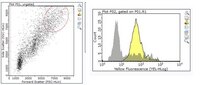
Flow Cytometry Analysis: A representative lot detected Thy 1.1 (CD90) in CAK1.3 and CAK4.4 cell lines (Ogasawara, K., et. al. (1990). Microbiol. Immunol. 34(12):1025-39).
Biological Information
Immunogen
Xenogenic rat thymocytes.
Epitope
extracellular domain
Clone
T11D7e
Concentration
Please refer to lot specific datasheet.
Host
Mouse
Specificity
Clone T11D7e detects Thy-1.1 (CD90) in rat thymocytes.
Flow Cytometry Analysis: 1 µg of this antibody detected Thy 1.1 (CD90) in one million rat thymocytes.
Usage Statement
Unless otherwise stated in our catalog or other company documentation accompanying the product(s), our products are intended for research use only and are not to be used for any other purpose, which includes but is not limited to, unauthorized commercial uses, in vitro diagnostic uses, ex vivo or in vivo therapeutic uses or any type of consumption or application to humans or animals.


ethyl acetate[814635_(Ethoxyethoxy)ethyl acetate-ALL].jpg)