Wenn Sie das Fenster schließen, wird Ihre Konfiguration nicht gespeichert, es sei denn, Sie haben Ihren Artikel in die Bestellung aufgenommen oder zu Ihren Favoriten hinzugefügt.
Klicken Sie auf OK, um das MILLIPLEX® MAP-Tool zu schließen oder auf Abbrechen, um zu Ihrer Auswahl zurückzukehren.
Wählen Sie konfigurierbare Panels & Premixed-Kits - ODER - Kits für die zelluläre Signaltransduktion & MAPmates™
Konfigurieren Sie Ihre MILLIPLEX® MAP-Kits und lassen sich den Preis anzeigen.
Konfigurierbare Panels & Premixed-Kits
Unser breites Angebot enthält Multiplex-Panels, für die Sie die Analyten auswählen können, die am besten für Ihre Anwendung geeignet sind. Unter einem separaten Register können Sie das Premixed-Cytokin-Format oder ein Singleplex-Kit wählen.
Kits für die zelluläre Signaltransduktion & MAPmates™
Wählen Sie gebrauchsfertige Kits zur Erforschung gesamter Signalwege oder Prozesse. Oder konfigurieren Sie Ihre eigenen Kits mit Singleplex MAPmates™.
Die folgenden MAPmates™ sollten nicht zusammen analysiert werden: -MAPmates™, die einen unterschiedlichen Assaypuffer erfordern. -Phosphospezifische und MAPmate™ Gesamtkombinationen wie Gesamt-GSK3β und Gesamt-GSK3β (Ser 9). -PanTyr und locusspezifische MAPmates™, z.B. Phospho-EGF-Rezeptor und Phospho-STAT1 (Tyr701). -Mehr als 1 Phospho-MAPmate™ für ein einziges Target (Akt, STAT3). -GAPDH und β-Tubulin können nicht mit Kits oder MAPmates™, die panTyr enthalten, analysiert werden.
.
Bestellnummer
Bestellinformationen
St./Pkg.
Liste
Dieser Artikel wurde zu Ihren Favoriten hinzugefügt.
Wählen Sie bitte Spezies, Panelart, Kit oder Probenart
Um Ihr MILLIPLEX® MAP-Kit zu konfigurieren, wählen Sie zunächst eine Spezies, eine Panelart und/oder ein Kit.
Custom Premix Selecting "Custom Premix" option means that all of the beads you have chosen will be premixed in manufacturing before the kit is sent to you.
Catalogue Number
Ordering Description
Qty/Pack
List
Dieser Artikel wurde zu Ihren Favoriten hinzugefügt.
Spezies
Panelart
Gewähltes Kit
Menge
Bestellnummer
Bestellinformationen
St./Pkg.
Listenpreis
96-Well Plate
Menge
Bestellnummer
Bestellinformationen
St./Pkg.
Listenpreis
Weitere Reagenzien hinzufügen (MAPmates erfordern die Verwendung eines Puffer- und Detektionskits)
Menge
Bestellnummer
Bestellinformationen
St./Pkg.
Listenpreis
48-602MAG
Buffer Detection Kit for Magnetic Beads
1 Kit
Platzsparende Option Kunden, die mehrere Kits kaufen, können ihre Multiplex-Assaykomponenten in Kunststoffbeuteln anstelle von Packungen erhalten, um eine kompaktere Lagerung zu ermöglichen.
Dieser Artikel wurde zu Ihren Favoriten hinzugefügt.
Das Produkt wurde in Ihre Bestellung aufgenommen
Sie können nun ein weiteres Kit konfigurieren, ein Premixed-Kit wählen, zur Kasse gehen oder das Bestell-Tool schließen.
This mouse monoclonal Anti-SUN2 Antibody, clone 3.1E, Cat. No. MABT880, is validated for use in Immunocytochemistry and Western Blotting for the detection of SUN2.
More>>This mouse monoclonal Anti-SUN2 Antibody, clone 3.1E, Cat. No. MABT880, is validated for use in Immunocytochemistry and Western Blotting for the detection of SUN2. Less<<
Anti-SUN2 Antibody, clone 3.1E: SDB (Sicherheitsdatenblätter), Analysenzertifikate und Qualitätszertifikate, Dossiers, Broschüren und andere verfügbare Dokumente.
SUN domain-containing protein 2 (UniProt Q9UH99; also known as Protein unc-84 homolog B, Rab5-interacting protein, Rab5IP, Sad1/unc-84 protein-like 2) is encoded by the SUN2 (also known as FRIGG, KIAA0668, RAB5IP, UNC84B) gene (Gene ID 25777) in human. It is a single-pass nuclear envelope transmembrane protein that is highly expressed in heart, lung, and muscle. It has a predicted transmembrane domain and a C-terminal region with similarity to the S. pombe spindle pole body protein Sad1. It may also facilitate a nuclear-centrosomal interaction required for nuclear migration and anchorage. It anchors chromosome movement in the prophase of meiosis and is required for telomere attachment to nuclear envelope and gametogenesis. SUN2 protein may also function on endocytic vesicles as a receptor for RAB5-GDP and participate in the activation of RAB5. Slight overexpression of SUN2 protein is seen in heart tissues form patients with congenital heart defects.
References
Product Information
Format
Purified
Presentation
Purified mouse IgG1κ in buffer containing 0.1 M Tris-Glycine (pH 7.4), 150 mM NaCl with 0.05% sodium azide.
This mouse monoclonal Anti-SUN2 Antibody, clone 3.1E, Cat. No. MABT880, is validated for use in Immunocytochemistry and Western Blotting for the detection of SUN2.
Key Applications
Immunocytochemistry
Western Blotting
Application Notes
Western Blotting Analysis: 0.5 µg/mL from a representative lot detected SUN2 in 10 µg of HepG2 cell lysate.
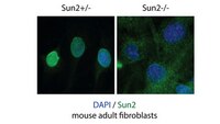
Immunocytochemistry Analysis: A representative lot immunostained nucleus of fibroblasts from Sun2+/-, but not Sun2-/- mice (Courtesy of Dr Brian Burke, Institute of Medical Biology, A*STAR).
Western Blotting Analysis: A representative lot detected SUN2 in heart tissue lysate from Sun2+/-, but not Sun2-/- mice (Courtesy of Dr Brian Burke, Institute of Medical Biology, A*STAR).
Immunofluorescence Analysis (IF): A representative lot detected mouse testis tissue (Courtesy of Dr Brian Burke, Institute of Medical Biology, A*STAR).
Immunofluorescence Analysis (IF): A representative lot tested on different mouse tissues (Courtesy of Dr Brian Burke, Institute of Medical Biology, A*STAR).
Immunofluorescence Analysis (IF): A representative lot detected immortalized fibroblasts derived from SUN2 +/+, SUN2 -/- , Sun1 -/- double knockout Sun2 -/- Sun1 -/- mouse pups (Courtesy of Dr Brian Burke, Institute of Medical Biology, A*STAR).
Biological Information
Immunogen
GST-tagged recombinant human SUN2 N-terminal LMNA-binding region fragment.
Clone
3.1E
Concentration
Please refer to lot specific datasheet.
Host
Mouse
Specificity
Target specificity of clone 3.1E was verified by immunocytochemistry and Western blotting analyses using fibroblasts and heart tissue samples from Sun2-knockout mice.
~80 kDa observed. 80.31/82.50/79.61 kDa (isoform 1/2/3) calculated. Uncharacterized bands may be observed in some lysate(s).
Physicochemical Information
Dimensions
Materials Information
Toxicological Information
Safety Information according to GHS
Safety Information
Product Usage Statements
Quality Assurance
Evaluated by Western Blotting in HeLa cell lysate.
Western Blotting Analysis: 0.5 µg/mL of this antibody detected SUN2 in 10 µg of HeLa cell lysate.
Usage Statement
Unless otherwise stated in our catalog or other company documentation accompanying the product(s), our products are intended for research use only and are not to be used for any other purpose, which includes but is not limited to, unauthorized commercial uses, in vitro diagnostic uses, ex vivo or in vivo therapeutic uses or any type of consumption or application to humans or animals.