Wenn Sie das Fenster schließen, wird Ihre Konfiguration nicht gespeichert, es sei denn, Sie haben Ihren Artikel in die Bestellung aufgenommen oder zu Ihren Favoriten hinzugefügt.
Klicken Sie auf OK, um das MILLIPLEX® MAP-Tool zu schließen oder auf Abbrechen, um zu Ihrer Auswahl zurückzukehren.
Wählen Sie konfigurierbare Panels & Premixed-Kits - ODER - Kits für die zelluläre Signaltransduktion & MAPmates™
Konfigurieren Sie Ihre MILLIPLEX® MAP-Kits und lassen sich den Preis anzeigen.
Konfigurierbare Panels & Premixed-Kits
Unser breites Angebot enthält Multiplex-Panels, für die Sie die Analyten auswählen können, die am besten für Ihre Anwendung geeignet sind. Unter einem separaten Register können Sie das Premixed-Cytokin-Format oder ein Singleplex-Kit wählen.
Kits für die zelluläre Signaltransduktion & MAPmates™
Wählen Sie gebrauchsfertige Kits zur Erforschung gesamter Signalwege oder Prozesse. Oder konfigurieren Sie Ihre eigenen Kits mit Singleplex MAPmates™.
Die folgenden MAPmates™ sollten nicht zusammen analysiert werden: -MAPmates™, die einen unterschiedlichen Assaypuffer erfordern. -Phosphospezifische und MAPmate™ Gesamtkombinationen wie Gesamt-GSK3β und Gesamt-GSK3β (Ser 9). -PanTyr und locusspezifische MAPmates™, z.B. Phospho-EGF-Rezeptor und Phospho-STAT1 (Tyr701). -Mehr als 1 Phospho-MAPmate™ für ein einziges Target (Akt, STAT3). -GAPDH und β-Tubulin können nicht mit Kits oder MAPmates™, die panTyr enthalten, analysiert werden.
.
Bestellnummer
Bestellinformationen
St./Pkg.
Liste
Dieser Artikel wurde zu Ihren Favoriten hinzugefügt.
Wählen Sie bitte Spezies, Panelart, Kit oder Probenart
Um Ihr MILLIPLEX® MAP-Kit zu konfigurieren, wählen Sie zunächst eine Spezies, eine Panelart und/oder ein Kit.
Custom Premix Selecting "Custom Premix" option means that all of the beads you have chosen will be premixed in manufacturing before the kit is sent to you.
Catalogue Number
Ordering Description
Qty/Pack
List
Dieser Artikel wurde zu Ihren Favoriten hinzugefügt.
Spezies
Panelart
Gewähltes Kit
Menge
Bestellnummer
Bestellinformationen
St./Pkg.
Listenpreis
96-Well Plate
Menge
Bestellnummer
Bestellinformationen
St./Pkg.
Listenpreis
Weitere Reagenzien hinzufügen (MAPmates erfordern die Verwendung eines Puffer- und Detektionskits)
Menge
Bestellnummer
Bestellinformationen
St./Pkg.
Listenpreis
48-602MAG
Buffer Detection Kit for Magnetic Beads
1 Kit
Platzsparende Option Kunden, die mehrere Kits kaufen, können ihre Multiplex-Assaykomponenten in Kunststoffbeuteln anstelle von Packungen erhalten, um eine kompaktere Lagerung zu ermöglichen.
Dieser Artikel wurde zu Ihren Favoriten hinzugefügt.
Das Produkt wurde in Ihre Bestellung aufgenommen
Sie können nun ein weiteres Kit konfigurieren, ein Premixed-Kit wählen, zur Kasse gehen oder das Bestell-Tool schließen.
Anti-STAT3 Rabbit pAb: SDB (Sicherheitsdatenblätter), Analysenzertifikate und Qualitätszertifikate, Dossiers, Broschüren und andere verfügbare Dokumente.
Synonyme: Anti-Signal Transducer and Activator of Transcription 3
Empfohlene Produkte
Übersicht
Replacement Information
Key Spec Table
Host
Rb
Description
Overview
This product has been discontinued.
We are offering Anti-Stat3 Antibody (Cat. No. 06-596) as a possible alternative. Please read the alternative product documentation carefully and contact technical service if you need additional information.
Recognizes native and denatured STAT3.
Catalogue Number
569388
Brand Family
Calbiochem®
Synonyms
Anti-Signal Transducer and Activator of Transcription 3
Application Data
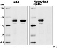
Detection of human STAT3 by immunoblotting. Samples: Whole cell lysate from HeLa cell treated with IFN-α (lane 1) or left untreated (lane 2). Primary antibody: Anti-STAT3 Rabbit pAb (Cat. No. 569388) (1:1000). Detection: chemiluminescence.
References
References
David, M., et al. 1995. Science269, 1721. Ihle, J.N. 1995. Nature377, 591. Wen, Z., et al. 1995. Cell82, 241. Darnell, J.E., Jr., et al. 1994. Science 264, 1415. Ihle, J.N., et al. 1994. Trends Biochem. Sci.19, 222.
Product Information
Form
Liquid
Formulation
In 150 mM NaCl, 10 mM HEPES, 50% glycerol, 0.01% BSA, pH 7.5.
For paraffin sections, pre-treatment in citrate buffer, pH 6.0 is required. Detects total STAT3 (phosphorylation-state independent) levels. Recognizes both native and denatured STAT3. Variables associated with assay conditions will dictate the proper working dilution.
Recommended Protocol for Immunoblotting
Solutions and Reagents • Transfer Buffer: 25 mM Tris base, 0.2 M glycine, 20% methanol, pH 8.5. • SDS Sample Buffer: 62.5 mM Tris-HCl, pH 6.8, 2% SDS, 10% glycerol, 50 mM DTT, 0.1% bromphenol blue. • 10X TBS (Tris-buffered saline): To prepare 1 liter, 24.2 g Tris base, 80 g NaCl, adjust pH to 7.6 with HCl. Dilute 1:10 for use. • Blocking Buffer: 1X TBS, 0.1% Tween®-20 detergent with 5% non-fat dry milk. • Primary Antibody Dilution Buffer: 1X TBS, 0.1% Tween®-20 detergent with 5% BSA • Wash Buffer (TBST): 1X TBS, 0.1% Tween®-20 detergent
Blotting Membrane Nitrocellulose or PVDF membranes may be used.
Protein Blotting A general protocol for sample preparation using 2x10⁶ SK-N-MC cells per well in a 6-well plate is as follows:
1. Culture cells in medium containing 0.5% FBS for 2 days. We recommend plating cells directly in 0.5% FBS media to reduce basal levels of STAT3 phosphorylation. 2. Aspirate media. Add fresh media without FBS. Culture for 2 h. Note: If cells are grown at high density, changing media before treating cells with regulator reduces basal STAT3 phosphorylation due to factors secreted by cells. 3. Aspirate media. Treat cells by adding fresh media containing regulator for desired time. 4. Aspirate media from cultures; wash cells with PBS; aspirate. 5. Lyse cells by adding 100 µl SDS Sample Buffer and immediately scrape the cells off the plate and transfer the extract to a microfuge tube. Keep on ice. 6. Sonicate for 2 s to shear DNA and reduce sample viscosity. 7. Heat sample to 95-100°C for 5 min. Cool on ice. 8. Microcentrifuge for 5 min. 9. Load 20 µl onto SDS-PAGE gel (10 cm x 10 cm). 10. Electrotransfer to nitrocellulose membrane.
As controls, we recommend using 10 µl of SK-N-MC cell extracts.
Membrane Blocking, Gel and Antibody Incubations 1. After transfer, wash membrane with 25 ml TBS for 5 min at room temperature. 2. Incubate membrane in 25 ml Blocking Buffer for 1-3 h at room temperature or overnight at 4°C. 3. Wash 3 times for 5 min each with 15 ml TBST. 4. Incubate membrane and primary antibody (at the appropriate dilution) in 10 ml Primary Antibody Dilution Buffer with gentle agitation overnight at 4°C. 5. Wash 3 times for 5 min each with 15 ml TBST. 6. Incubate membrane with conjugated secondary antibody at the appropriate dilution in 10 ml Blocking Buffer with gentle agitation for 1 h at room temperature. 7. Wash membrane as in step 5.
Detection of Proteins Chemiluminescence.
Biological Information
Immunogen
a synthetic peptide corresponding to amino acids surrounding Tyr⁷⁰⁵ of mouse STAT3
Immunogen
Mouse
Host
Rabbit
Isotype
IgG
Physicochemical Information
Dimensions
Materials Information
Toxicological Information
Safety Information according to GHS
Safety Information
Product Usage Statements
Storage and Shipping Information
Ship Code
Blue Ice Only
Toxicity
Standard Handling
Storage
-20°C
Avoid freeze/thaw
Avoid freeze/thaw
Do not freeze
Ok to freeze
Special Instructions
Following initial thaw, aliquot and freeze (-20°C).
Packaging Information
Transport Information
Supplemental Information
Specifications
Global Trade Item Number
Bestellnummer
GTIN
569388
0
Documentation
Anti-STAT3 Rabbit pAb Analysenzertifikate
Titel
Chargennummer
569388
Literatur
Übersicht
David, M., et al. 1995. Science269, 1721. Ihle, J.N. 1995. Nature377, 591. Wen, Z., et al. 1995. Cell82, 241. Darnell, J.E., Jr., et al. 1994. Science 264, 1415. Ihle, J.N., et al. 1994. Trends Biochem. Sci.19, 222.
Datenblatt
Note that this data sheet is not lot-specific and is representative of the current specifications for this product. Please consult the vial label and the certificate of analysis for information on specific lots. Also note that shipping conditions may differ from storage conditions.
Revision
06-July-2010 JSW
Synonyms
Anti-Signal Transducer and Activator of Transcription 3
Detection of human STAT3 by immunoblotting. Samples: Whole cell lysate from HeLa cell treated with IFN-α (lane 1) or left untreated (lane 2). Primary antibody: Anti-STAT3 Rabbit pAb (Cat. No. 569388) (1:1000). Detection: chemiluminescence.
Description
Protein A and immunoaffinity purified rabbit polyclonal antibody. Recognizes the ~90 kDa STAT3 protein.
Background
STAT3 is activated by a variety of cytokines including interleukins, CNTF and EPO. Activation of STAT3 is accompanied by tyrosine phosphorylation at Tyr705 which induces dimerization, nuclear translocation and DNA binding. Transcriptional activation also appears to be regulated by serine phosphorylation (Ser727) probably via MAP-like kinases. As phosphorylation of STAT3 at Tyr705 is essential for dimerization and DNA binding, phosphorylation at this site is an excellent marker of STAT3 activity.
Host
Rabbit
Immunogen species
Mouse
Immunogen
a synthetic peptide corresponding to amino acids surrounding Tyr⁷⁰⁵ of mouse STAT3
Isotype
IgG
Species
human, mouse, rat
Form
Liquid
Formulation
In 150 mM NaCl, 10 mM HEPES, 50% glycerol, 0.01% BSA, pH 7.5.
Preservative
None
Comments
For paraffin sections, pre-treatment in citrate buffer, pH 6.0 is required. Detects total STAT3 (phosphorylation-state independent) levels. Recognizes both native and denatured STAT3. Variables associated with assay conditions will dictate the proper working dilution.
Recommended Protocol for Immunoblotting
Solutions and Reagents • Transfer Buffer: 25 mM Tris base, 0.2 M glycine, 20% methanol, pH 8.5. • SDS Sample Buffer: 62.5 mM Tris-HCl, pH 6.8, 2% SDS, 10% glycerol, 50 mM DTT, 0.1% bromphenol blue. • 10X TBS (Tris-buffered saline): To prepare 1 liter, 24.2 g Tris base, 80 g NaCl, adjust pH to 7.6 with HCl. Dilute 1:10 for use. • Blocking Buffer: 1X TBS, 0.1% Tween®-20 detergent with 5% non-fat dry milk. • Primary Antibody Dilution Buffer: 1X TBS, 0.1% Tween®-20 detergent with 5% BSA • Wash Buffer (TBST): 1X TBS, 0.1% Tween®-20 detergent
Blotting Membrane Nitrocellulose or PVDF membranes may be used.
Protein Blotting A general protocol for sample preparation using 2x10⁶ SK-N-MC cells per well in a 6-well plate is as follows:
1. Culture cells in medium containing 0.5% FBS for 2 days. We recommend plating cells directly in 0.5% FBS media to reduce basal levels of STAT3 phosphorylation. 2. Aspirate media. Add fresh media without FBS. Culture for 2 h. Note: If cells are grown at high density, changing media before treating cells with regulator reduces basal STAT3 phosphorylation due to factors secreted by cells. 3. Aspirate media. Treat cells by adding fresh media containing regulator for desired time. 4. Aspirate media from cultures; wash cells with PBS; aspirate. 5. Lyse cells by adding 100 µl SDS Sample Buffer and immediately scrape the cells off the plate and transfer the extract to a microfuge tube. Keep on ice. 6. Sonicate for 2 s to shear DNA and reduce sample viscosity. 7. Heat sample to 95-100°C for 5 min. Cool on ice. 8. Microcentrifuge for 5 min. 9. Load 20 µl onto SDS-PAGE gel (10 cm x 10 cm). 10. Electrotransfer to nitrocellulose membrane.
As controls, we recommend using 10 µl of SK-N-MC cell extracts.
Membrane Blocking, Gel and Antibody Incubations 1. After transfer, wash membrane with 25 ml TBS for 5 min at room temperature. 2. Incubate membrane in 25 ml Blocking Buffer for 1-3 h at room temperature or overnight at 4°C. 3. Wash 3 times for 5 min each with 15 ml TBST. 4. Incubate membrane and primary antibody (at the appropriate dilution) in 10 ml Primary Antibody Dilution Buffer with gentle agitation overnight at 4°C. 5. Wash 3 times for 5 min each with 15 ml TBST. 6. Incubate membrane with conjugated secondary antibody at the appropriate dilution in 10 ml Blocking Buffer with gentle agitation for 1 h at room temperature. 7. Wash membrane as in step 5.
Detection of Proteins Chemiluminescence.
Storage
Avoid freeze/thaw
-20°C
Do Not Freeze
Ok to freeze
Special Instructions
Following initial thaw, aliquot and freeze (-20°C).
Toxicity
Standard Handling
References
David, M., et al. 1995. Science269, 1721. Ihle, J.N. 1995. Nature377, 591. Wen, Z., et al. 1995. Cell82, 241. Darnell, J.E., Jr., et al. 1994. Science 264, 1415. Ihle, J.N., et al. 1994. Trends Biochem. Sci.19, 222.