Wenn Sie das Fenster schließen, wird Ihre Konfiguration nicht gespeichert, es sei denn, Sie haben Ihren Artikel in die Bestellung aufgenommen oder zu Ihren Favoriten hinzugefügt.
Klicken Sie auf OK, um das MILLIPLEX® MAP-Tool zu schließen oder auf Abbrechen, um zu Ihrer Auswahl zurückzukehren.
Wählen Sie konfigurierbare Panels & Premixed-Kits - ODER - Kits für die zelluläre Signaltransduktion & MAPmates™
Konfigurieren Sie Ihre MILLIPLEX® MAP-Kits und lassen sich den Preis anzeigen.
Konfigurierbare Panels & Premixed-Kits
Unser breites Angebot enthält Multiplex-Panels, für die Sie die Analyten auswählen können, die am besten für Ihre Anwendung geeignet sind. Unter einem separaten Register können Sie das Premixed-Cytokin-Format oder ein Singleplex-Kit wählen.
Kits für die zelluläre Signaltransduktion & MAPmates™
Wählen Sie gebrauchsfertige Kits zur Erforschung gesamter Signalwege oder Prozesse. Oder konfigurieren Sie Ihre eigenen Kits mit Singleplex MAPmates™.
Die folgenden MAPmates™ sollten nicht zusammen analysiert werden: -MAPmates™, die einen unterschiedlichen Assaypuffer erfordern. -Phosphospezifische und MAPmate™ Gesamtkombinationen wie Gesamt-GSK3β und Gesamt-GSK3β (Ser 9). -PanTyr und locusspezifische MAPmates™, z.B. Phospho-EGF-Rezeptor und Phospho-STAT1 (Tyr701). -Mehr als 1 Phospho-MAPmate™ für ein einziges Target (Akt, STAT3). -GAPDH und β-Tubulin können nicht mit Kits oder MAPmates™, die panTyr enthalten, analysiert werden.
.
Bestellnummer
Bestellinformationen
St./Pkg.
Liste
Dieser Artikel wurde zu Ihren Favoriten hinzugefügt.
Wählen Sie bitte Spezies, Panelart, Kit oder Probenart
Um Ihr MILLIPLEX® MAP-Kit zu konfigurieren, wählen Sie zunächst eine Spezies, eine Panelart und/oder ein Kit.
Custom Premix Selecting "Custom Premix" option means that all of the beads you have chosen will be premixed in manufacturing before the kit is sent to you.
Catalogue Number
Ordering Description
Qty/Pack
List
Dieser Artikel wurde zu Ihren Favoriten hinzugefügt.
Spezies
Panelart
Gewähltes Kit
Menge
Bestellnummer
Bestellinformationen
St./Pkg.
Listenpreis
96-Well Plate
Menge
Bestellnummer
Bestellinformationen
St./Pkg.
Listenpreis
Weitere Reagenzien hinzufügen (MAPmates erfordern die Verwendung eines Puffer- und Detektionskits)
Menge
Bestellnummer
Bestellinformationen
St./Pkg.
Listenpreis
48-602MAG
Buffer Detection Kit for Magnetic Beads
1 Kit
Platzsparende Option Kunden, die mehrere Kits kaufen, können ihre Multiplex-Assaykomponenten in Kunststoffbeuteln anstelle von Packungen erhalten, um eine kompaktere Lagerung zu ermöglichen.
Dieser Artikel wurde zu Ihren Favoriten hinzugefügt.
Das Produkt wurde in Ihre Bestellung aufgenommen
Sie können nun ein weiteres Kit konfigurieren, ein Premixed-Kit wählen, zur Kasse gehen oder das Bestell-Tool schließen.
Anti-SIRP alpha, clone SAF10.1, Cat. No. MABS2188, is a mouse monoclonal antibody that detects Tyrosine-protein phosphatase non-receptor type substrate 1 (SIRP alpha) and has been tested for use in ELISA and Western Blotting.
More>>Anti-SIRP alpha, clone SAF10.1, Cat. No. MABS2188, is a mouse monoclonal antibody that detects Tyrosine-protein phosphatase non-receptor type substrate 1 (SIRP alpha) and has been tested for use in ELISA and Western Blotting. Less<<
Anti-SIRP alpha Antibody, clone SAF10.1: SDB (Sicherheitsdatenblätter), Analysenzertifikate und Qualitätszertifikate, Dossiers, Broschüren und andere verfügbare Dokumente.
Tyrosine-protein phosphatase non-receptor type substrate 1
SHP substrate 1
SHPS-1
Brain Ig-like molecule with tyrosine-based activation motifs
Bit
CD172 antigen-like family member A
Inhibitory receptor SHPS-1
Macrophage fusion receptor
MyD-1 antigen
Signal-regulatory protein alpha-1
Sirp-alpha-1
Signal-regulatory protein alpha-2
Sirp-alpha-2
Signal-regulatory protein alpha-3
Sirp-alpha-3
p84
CD172a
Background Information
Tyrosine-protein phosphatase non-receptor type substrate 1 (UniProt: P78324; also known as SHP substrate 1, SHPS-1, Brain Ig-like molecule with tyrosine-based activation motifs, Bit, CD172 antigen-like family member A, Inhibitory receptor SHPS-1, Macrophage fusion receptor, MyD-1 antigen, Signal-regulatory protein alpha-1, Sirp-alpha-1, Signal-regulatory protein alpha-2, Sirp-alpha-2, Signal-regulatory protein alpha-3, Sirp-alpha-3, p84, CD172a) is encoded by the SIRPA (also known as BIT, MFR, MYD1, PTPNS1, SHPS1, SIRP) gene (Gene ID: 140885) in human. SIRP alpha is a single-pass type I membrane protein of the Ig superfamily that is ubiquitously expressed with high expression reported in brain. It is also detected on myeloid cells, but not on T cells. SIRP alpha is synthesized with a signal peptide (aa 1-30), which is subsequently cleaved off to generate the mature form that contains an extracellular domain (aa 31-373), a transmembrane domain (aa 374-394) and a cytoplasmic domain (aa 395-504). The extracellular region contains three Ig-like loops. The most distal loop (D1) contains and Ig-like V type domain (aa 32-137) whereas the two membrane proximal loops contain Ig-like C1 type domains (aa 148-247 and 254-348). SIRP alpha serves as an immunoglobulin-like cell surface receptor for CD47 and their interactions is reported to mediate negative regulation of several monocyte/ macrophage functions. CD47 binding prevents maturation of immature dendritic cells and inhibits cytokine production by mature dendritic cells. SIRP alpha also acts as a docking protein and induces translocation of PTPN6, PTPN11, and other binding partners from the cytosol to the plasma membrane. SIRP alpha is phosphorylated on tyrosine residues in response to stimulation with EGF, growth hormone, insulin, and platelet derived growth factor. Clone SAF10.1 recognizes an epitope in the membrane most distal domain, termed as the D1 domain. (Ref.: Lee, WY et al., (2007). J Immunol.; 179(11); 7741-7750).
References
Product Information
Format
Purified
Presentation
Purified mouse monoclonal antibody IgG1 in buffer containing 0.1 M Tris-Glycine (pH 7.4), 150 mM NaCl with 0.05% sodium azide.
Applications
Application
Anti-SIRP alpha, clone SAF10.1, Cat. No. MABS2188, is a mouse monoclonal antibody that detects Tyrosine-protein phosphatase non-receptor type substrate 1 (SIRP alpha) and has been tested for use in ELISA and Western Blotting.
Key Applications
ELISA
Western Blotting
Application Notes
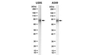
Western Blotting Analysis: 1 µg/mL from a representative lot detected SIRP alpha in A549 cell lysate.
Western Blotting Analysis: A represetntative lot detected SIRP alpha in Western Blotting applications (Lee, W.Y., et. al. (2007). J. Immunol. 179(11); 7741-50).
ELISA Analysis: A represetntative lot detected SIRP alpha in ELISA applications (Lee, W.Y., et. al. (2007). J. Immunol. 179(11); 7741-50).
Biological Information
Immunogen
Eukaryotically expressed SIRP-Fc fusion protein consisting of the extracellular domain.
Clone
SAF10.1
Concentration
Please refer to lot specific datasheet.
Host
Mouse
Specificity
Clone SAF10.1 is a mouse monoclonal antibody that detects human Tyrosine-protein phosphatase non-receptor type substrate 1 (SIRP alpha). It targets an epitope in the membrane most distal domain (D1).
~75 kDa observed; 54.97 kDa calculated. Uncharacterized bands may be observed in some lysate(s).
Physicochemical Information
Dimensions
Materials Information
Toxicological Information
Safety Information according to GHS
Safety Information
Product Usage Statements
Quality Assurance
Evaluated by Western Blotting in U20S cell lysate.
Western Blotting Analysis: 1 µg/mL of this antibody detected SIRP alpha in U20S cell lysate.
Usage Statement
Unless otherwise stated in our catalog or other company documentation accompanying the product(s), our products are intended for research use only and are not to be used for any other purpose, which includes but is not limited to, unauthorized commercial uses, in vitro diagnostic uses, ex vivo or in vivo therapeutic uses or any type of consumption or application to humans or animals.
Signal regulatory proteins (SIRP-alpha, -beta, and -gamma) are important regulators of several innate immune functions that include leukocyte migration. Membrane distal (D1) domains of SIRPalpha and SIRPgamma, but not SIRPbeta, mediate binding to a cellular ligand termed CD47. Because the extracellular domains of all SIRPs are highly homologous, we hypothesized that some of the 16 residues unique to SIRPalpha.D1 mediate binding to CD47. By site-directed mutagenesis, we determined that SIRPalpha binding to CD47 is independent of N-glycosylation. We also identified three residues critical for CD47 binding by exchanging residues on SIRPalpha with corresponding residues from SIRPbeta. Cumulative substitutions of the critical residues into SIRPbeta resulted in de novo binding of the mutant protein to CD47. Homology modeling of SIRPalpha.D1 revealed topological relationships among critical residues and allowed the identification of critical residues common to SIRPalpha and SIRPbeta. Mapping these critical residues onto the recently reported crystal structure of SIRPalpha.D1 revealed a novel region that is required for CD47 binding and is distinct and lateral to another putative CD47 binding site described on that crystal structure. The importance of this lateral region in mediating SIRPalpha.D1 binding to CD47 was confirmed by epitope mapping analyses of anti-SIRP Abs. These observations highlight a complex nature of the ligand binding requirements for SIRPalpha that appear to be dependent on two distinct but adjacent regions on the membrane distal Ig loop. A better understanding of the structural basis of SIRPalpha/CD47 interactions may provide insights into therapeutics targeting pathologic inflammation.