Wenn Sie das Fenster schließen, wird Ihre Konfiguration nicht gespeichert, es sei denn, Sie haben Ihren Artikel in die Bestellung aufgenommen oder zu Ihren Favoriten hinzugefügt.
Klicken Sie auf OK, um das MILLIPLEX® MAP-Tool zu schließen oder auf Abbrechen, um zu Ihrer Auswahl zurückzukehren.
Wählen Sie konfigurierbare Panels & Premixed-Kits - ODER - Kits für die zelluläre Signaltransduktion & MAPmates™
Konfigurieren Sie Ihre MILLIPLEX® MAP-Kits und lassen sich den Preis anzeigen.
Konfigurierbare Panels & Premixed-Kits
Unser breites Angebot enthält Multiplex-Panels, für die Sie die Analyten auswählen können, die am besten für Ihre Anwendung geeignet sind. Unter einem separaten Register können Sie das Premixed-Cytokin-Format oder ein Singleplex-Kit wählen.
Kits für die zelluläre Signaltransduktion & MAPmates™
Wählen Sie gebrauchsfertige Kits zur Erforschung gesamter Signalwege oder Prozesse. Oder konfigurieren Sie Ihre eigenen Kits mit Singleplex MAPmates™.
Die folgenden MAPmates™ sollten nicht zusammen analysiert werden: -MAPmates™, die einen unterschiedlichen Assaypuffer erfordern. -Phosphospezifische und MAPmate™ Gesamtkombinationen wie Gesamt-GSK3β und Gesamt-GSK3β (Ser 9). -PanTyr und locusspezifische MAPmates™, z.B. Phospho-EGF-Rezeptor und Phospho-STAT1 (Tyr701). -Mehr als 1 Phospho-MAPmate™ für ein einziges Target (Akt, STAT3). -GAPDH und β-Tubulin können nicht mit Kits oder MAPmates™, die panTyr enthalten, analysiert werden.
.
Bestellnummer
Bestellinformationen
St./Pkg.
Liste
Dieser Artikel wurde zu Ihren Favoriten hinzugefügt.
Wählen Sie bitte Spezies, Panelart, Kit oder Probenart
Um Ihr MILLIPLEX® MAP-Kit zu konfigurieren, wählen Sie zunächst eine Spezies, eine Panelart und/oder ein Kit.
Custom Premix Selecting "Custom Premix" option means that all of the beads you have chosen will be premixed in manufacturing before the kit is sent to you.
Catalogue Number
Ordering Description
Qty/Pack
List
Dieser Artikel wurde zu Ihren Favoriten hinzugefügt.
Spezies
Panelart
Gewähltes Kit
Menge
Bestellnummer
Bestellinformationen
St./Pkg.
Listenpreis
96-Well Plate
Menge
Bestellnummer
Bestellinformationen
St./Pkg.
Listenpreis
Weitere Reagenzien hinzufügen (MAPmates erfordern die Verwendung eines Puffer- und Detektionskits)
Menge
Bestellnummer
Bestellinformationen
St./Pkg.
Listenpreis
48-602MAG
Buffer Detection Kit for Magnetic Beads
1 Kit
Platzsparende Option Kunden, die mehrere Kits kaufen, können ihre Multiplex-Assaykomponenten in Kunststoffbeuteln anstelle von Packungen erhalten, um eine kompaktere Lagerung zu ermöglichen.
Dieser Artikel wurde zu Ihren Favoriten hinzugefügt.
Das Produkt wurde in Ihre Bestellung aufgenommen
Sie können nun ein weiteres Kit konfigurieren, ein Premixed-Kit wählen, zur Kasse gehen oder das Bestell-Tool schließen.
Anti-Rai, clone IF15, Cat. No. MABN2286, is a mouse monoclonal antibody that detects SHC-transforming protein 3 (Rai) and is tested for use in Immunocytochemistry, Immunohistochemistry (Paraffin), and Western Blotting.
More>>Anti-Rai, clone IF15, Cat. No. MABN2286, is a mouse monoclonal antibody that detects SHC-transforming protein 3 (Rai) and is tested for use in Immunocytochemistry, Immunohistochemistry (Paraffin), and Western Blotting. Less<<
Src homology 2 domain-containing-transforming protein C3
SH2 domain protein C3
Background Information
SHC-transforming protein 3 (UniProt: Q92529; also known as Neuronal Shc, N-Shc, Protein Rai, SHC-transforming protein C, Src homology 2 domain-containing-transforming protein C3, SH2 domain protein C3, Rai) is encoded by the SHC3 (also known as NSHC, SHCC) gene (Gene ID:53358) in human. Rai protein is mainly expressed in the brain and its higher expression is observed in cerebral cortex, frontal and temporal lobes, hippocampus, and occipital pole. Its expression is also reported in the enteric glial cells in the human gut. It serves as a signaling adapter and is involved in the signal transduction pathways of neurotrophin-activated Trk receptors in cortical neurons. Rai also serves as a physiological substrate of the receptor tyrosine kinase Ret and increases Ret-dependent survival via PI3 kinase/Akt pathway activation. It also potentiates MAP kinase signaling. Rai activation also plays a role in long-term potential and is neuroprotective against certain ischemia and oxidative stress. Evolutionarily it correlates well with vertebrate brain development. Its reduced levels are observed in brain of Alzheimer s disease subjects. Two isoforms of Rai have been described (p64 and p52) that are produced by alternative splicing. (Ref.: Triaca, V., et al. (2018). Aging. 10(1); 5-6; Sagi, O., et al. (2015). Ageing Res. Rev. 19; 34-42; Villanacci, V., et al. (2008). Neurogastroenterol. Motil. 20(3); 206-212).
References
Product Information
Format
Purified
Presentation
Purified mouse monoclonal antibody in buffer containing 0.1 M Tris-Glycine (pH 7.4), 150 mM NaCl with 0.05% sodium azide.
Anti-Rai, clone IF15, Cat. No. MABN2286, is a mouse monoclonal antibody that detects SHC-transforming protein 3 (Rai) and is tested for use in Immunocytochemistry, Immunohistochemistry (Paraffin), and Western Blotting.
Key Applications
Western Blotting
Immunohistochemistry (Paraffin)
Immunocytochemistry
Application Notes
Tested Applications
Immunohistochemistry Applications: A representative lot detected Rai in Immunohistochemistry applications (Villanacci, V., et. al. (2008). Neurogastroenterol Motil. 20(3):206-12).
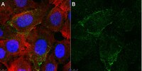
Immunocytochemistry Analysis: A 1:100 dilution from a representative lot detected Rai in U2OS cells.
Immunohistochemistry (Paraffin) Analysis: A 1:500 dilution from a representative lot detected Rai in human esophagus, stomach, and colon tissue sections.
Note: Actual optimal working dilutions must be determined by end user as specimens, and experimental conditions may vary with the end user
Biological Information
Immunogen
Recombinant fragment corresponding to 164 amino acids CH1 domain of human SHC-transforming protein 3 (Rai).
Epitope
C-terminal half
Clone
IF15-E2
Concentration
0.5 mg/mL. Please refer to guidance on suggested starting dilutions and/or titers per application and sample type.
Host
Mouse
Specificity
Clone IF15-E2 is a mouse monoclonal antibody that detects SHC-transforming protein 3/RAI. It targets an epitope within the CH1 domain from the C-terminal half.
~58 kDa observed; 64.06 kDa calculated. Uncharacterized bands may be observed in some lysate(s).
Physicochemical Information
Dimensions
Materials Information
Toxicological Information
Safety Information according to GHS
Safety Information
Product Usage Statements
Quality Assurance
Evaluated by Western Blotting in Human brain tissue lysate.
Western Blotting Analysis: A 1:500 dilution of this antibody detected Rai in Human brain tissue lysate.
Usage Statement
Unless otherwise stated in our catalog or other company documentation accompanying the product(s), our products are intended for research use only and are not to be used for any other purpose, which includes but is not limited to, unauthorized commercial uses, in vitro diagnostic uses, ex vivo or in vivo therapeutic uses or any type of consumption or application to humans or animals.
Storage and Shipping Information
Storage Conditions
Recommend storage at +2°C to +8°C. For long term storage antibodies can be kept at -20°C. Avoid repeated freeze-thaws.