Wenn Sie das Fenster schließen, wird Ihre Konfiguration nicht gespeichert, es sei denn, Sie haben Ihren Artikel in die Bestellung aufgenommen oder zu Ihren Favoriten hinzugefügt.
Klicken Sie auf OK, um das MILLIPLEX® MAP-Tool zu schließen oder auf Abbrechen, um zu Ihrer Auswahl zurückzukehren.
Wählen Sie konfigurierbare Panels & Premixed-Kits - ODER - Kits für die zelluläre Signaltransduktion & MAPmates™
Konfigurieren Sie Ihre MILLIPLEX® MAP-Kits und lassen sich den Preis anzeigen.
Konfigurierbare Panels & Premixed-Kits
Unser breites Angebot enthält Multiplex-Panels, für die Sie die Analyten auswählen können, die am besten für Ihre Anwendung geeignet sind. Unter einem separaten Register können Sie das Premixed-Cytokin-Format oder ein Singleplex-Kit wählen.
Kits für die zelluläre Signaltransduktion & MAPmates™
Wählen Sie gebrauchsfertige Kits zur Erforschung gesamter Signalwege oder Prozesse. Oder konfigurieren Sie Ihre eigenen Kits mit Singleplex MAPmates™.
Die folgenden MAPmates™ sollten nicht zusammen analysiert werden: -MAPmates™, die einen unterschiedlichen Assaypuffer erfordern. -Phosphospezifische und MAPmate™ Gesamtkombinationen wie Gesamt-GSK3β und Gesamt-GSK3β (Ser 9). -PanTyr und locusspezifische MAPmates™, z.B. Phospho-EGF-Rezeptor und Phospho-STAT1 (Tyr701). -Mehr als 1 Phospho-MAPmate™ für ein einziges Target (Akt, STAT3). -GAPDH und β-Tubulin können nicht mit Kits oder MAPmates™, die panTyr enthalten, analysiert werden.
.
Bestellnummer
Bestellinformationen
St./Pkg.
Liste
Dieser Artikel wurde zu Ihren Favoriten hinzugefügt.
Wählen Sie bitte Spezies, Panelart, Kit oder Probenart
Um Ihr MILLIPLEX® MAP-Kit zu konfigurieren, wählen Sie zunächst eine Spezies, eine Panelart und/oder ein Kit.
Custom Premix Selecting "Custom Premix" option means that all of the beads you have chosen will be premixed in manufacturing before the kit is sent to you.
Catalogue Number
Ordering Description
Qty/Pack
List
Dieser Artikel wurde zu Ihren Favoriten hinzugefügt.
Spezies
Panelart
Gewähltes Kit
Menge
Bestellnummer
Bestellinformationen
St./Pkg.
Listenpreis
96-Well Plate
Menge
Bestellnummer
Bestellinformationen
St./Pkg.
Listenpreis
Weitere Reagenzien hinzufügen (MAPmates erfordern die Verwendung eines Puffer- und Detektionskits)
Menge
Bestellnummer
Bestellinformationen
St./Pkg.
Listenpreis
48-602MAG
Buffer Detection Kit for Magnetic Beads
1 Kit
Platzsparende Option Kunden, die mehrere Kits kaufen, können ihre Multiplex-Assaykomponenten in Kunststoffbeuteln anstelle von Packungen erhalten, um eine kompaktere Lagerung zu ermöglichen.
Dieser Artikel wurde zu Ihren Favoriten hinzugefügt.
Das Produkt wurde in Ihre Bestellung aufgenommen
Sie können nun ein weiteres Kit konfigurieren, ein Premixed-Kit wählen, zur Kasse gehen oder das Bestell-Tool schließen.
ABN1657
Sigma-AldrichAnti-RME-8
Anti-RME-8, Cat. No. ABN1657, is a highly specific rabbit polyclonal antibody that targets RME-8 and has been tested in Immunocytochemistry and Western Blotting.
More>>Anti-RME-8, Cat. No. ABN1657, is a highly specific rabbit polyclonal antibody that targets RME-8 and has been tested in Immunocytochemistry and Western Blotting. Less<<
Anti-RME-8: SDB (Sicherheitsdatenblätter), Analysenzertifikate und Qualitätszertifikate, Dossiers, Broschüren und andere verfügbare Dokumente.
DnaJ homolog subfamily C member 13 (UniProt O75165; also known as Required for receptor-mediated endocytosis 8, RME-8) is encoded by the DNAJC13 (also known as KIAA0678, PARK21, RME8) gene (Gene ID 23317) in human. Originally identified as a C. elegans protein involved in mediating endocytosis, , receptor-mediated endocytosis 8 (RME-8) is a member of the DNAJ family of proteins implicated in regulating protein folding through associated chaperones. RME-8 is shown to interact with Hsc70/HSP-1 and sorting nexin 1 (SNX-1), loss of RME-8, SNX-1 or Hsc70 affects retrograde trafficking at the early endosomes-TGN interface. In addition, RME-8 is demonstrated to coordinate the function of the WASH complex and the retromer SNX dimer. The pentameric WASH complex is comprised of WASH1, strumpellin, SWIP (strumpellin- and wash-interacting protein), FAM21, and CCDC53. RME-8 N-terminal J region (a.a. 1-453) involved in membrane association is found to be involved in establishing interaction with the first third of FAM21 tail domain. WASH complex is responsible for the generation of branched actin networks on endosomes and the creation of microdomains into which specific cargoes can be concentrated. Loss of RME-8 function results in a profound endosomal tubulation, more extensive than the endosomal tubulation phenotype resulted from loss of the WASH complex.
References
Product Information
Format
Affinity Purified
Presentation
Purified rabbit polyclonal antibody in buffer containing PBS with 0.05% sodium azide and 50% glycerol.
Anti-RME-8, Cat. No. ABN1657, is a highly specific rabbit polyclonal antibody that targets RME-8 and has been tested in Immunocytochemistry and Western Blotting.
Key Applications
Immunocytochemistry
Western Blotting
Application Notes
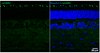
Immunocytochemistry Analysis: A 1:200 dilution from a representative lot detected shRNA-mediated RME-8 downregulation in COS-7 cells (Courtesy of Peter McPherson, Ph.D., FRSC, and Martine Girard, PhD, McGill University, Canada).
Western Blotting Analysis: A representative lot detected RME-8 associated with the 20,000 xg pelleted insoluble fraction from the HEPES buffer homogenates of human skin fibroblast MCH46, breast epithelial MCF10A, and embryonic kidney HEK293T cells (BT-474, HeLa, SK-BR-3) cells (Ioannou, M.S., et al. (2016). J. Biol. Chem. In press).
Western Blotting Analysis: A 1:1,000 dilution from a representative lot detected shRNA-mediated RME-8 downregulation in HEK293T cells (Courtesy of Peter McPherson, Ph.D., FRSC, and Martine Girard, PhD, McGill University, Canada).
Immunocytochemistry Analysis: A representative deteced a reduced RME-8 immunoreactivity in HeLa cells transfected with shRNA targeting either RME-8 or Spastin (Freeman, C.L., et al. (2014). J. Cell Sci. 127(Pt 9):2053-2070).
Immunocytochemistry Analysis: A representative lot detected puntate endosomal RME-8 immunoreactivity by fluorescent immunocytochemistry staining of 2% paraformaldehyde-fixed, 0.2% Triton X-100-permeabilized COS-7 cells. LIttle RME-8 immunoreactivity was seen associated with TGN, lysosome, or plasma membrane, RME-8 shRNA transfection greately abolished cellular immunostaining (Girard, M., et al. (2005). J. Biol. Chem. 280(48):40135-40143).
Western Blotting Analysis: A representative lot detected the presence of RME-8 in WASH complex immunoprecipitated from HeLa cells (BT-474, HeLa, SK-BR-3) cells (Freeman, C.L., et al. (2014). J. Cell Sci. 127(Pt 9):2053-2070).
Western Blotting Analysis: Representative lots detected a greatly reduced ~200 kDa target band in lysates from RME-8 shRNA-transfected monkey (COS-7) and human (BT-474, HeLa, SK-BR-3) cells (Freeman, C.L., et al. (2014). J. Cell Sci. 127(Pt 9):2053-2070; Girard, M., and McPherson, P.S. (2008). FEBS Lett. 582(6):961-966; Girard, M., et al. (2005). J. Biol. Chem. 280(48):40135-40143).
Western Blotting Analysis: A representative lot detected the ~200 kDa RME-8 target band in postnuclear extracts from multiple rat tissues as well as from human, monkey, mouse, and rat cell lines. RME-8 was found enriched in rat kidney tissue-derived subcellular fractions representing microsomes and clathrin-coated vesicles (CCVs), but not cytosol or plasma membrane (Girard, M., et al. (2005). J. Biol. Chem. 280(48):40135-40143).
Biological Information
Immunogen
KLH-conjugated linear peptide corresponding to the C-terminal end sequence of human RME-8.
Epitope
C-terminus
Concentration
Please refer to lot specific datasheet.
Host
Rabbit
Specificity
This rabbit polyclonal antibody detected shRNA-mediated cellular RME-8 knockwon by immunoblotting and fluorescent immunocytochemistry (Freeman, C.L., et al. (2014). J. Cell Sci. 127(Pt 9):2053-2070; Girard, M., and McPherson, P.S. (2008). FEBS Lett. 582(6):961-966; Girard, M., et al. J. Biol. Chem. 280(48):40135-40143).
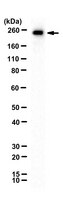
~220 kDa observed. 254.4 kDa calculated. Uncharacterized bands may be observed in some lysate(s).
Physicochemical Information
Dimensions
Materials Information
Toxicological Information
Safety Information according to GHS
Safety Information
Product Usage Statements
Quality Assurance
Evaluated by Western Blotting in NIH/3T3 cell lysate.
Western Blotting Analysis: A 1:500 dilution of this antibody detected RME-8 in 10 µg of NIH/3T3 cell lysate.
Usage Statement
Unless otherwise stated in our catalog or other company documentation accompanying the product(s), our products are intended for research use only and are not to be used for any other purpose, which includes but is not limited to, unauthorized commercial uses, in vitro diagnostic uses, ex vivo or in vivo therapeutic uses or any type of consumption or application to humans or animals.
Storage and Shipping Information
Storage Conditions
Stable for 1 year at -20°C from date of receipt. Note: Variability in freezer temperatures below -20°C may cause glycerol containing solutions to become frozen during storage.