Wenn Sie das Fenster schließen, wird Ihre Konfiguration nicht gespeichert, es sei denn, Sie haben Ihren Artikel in die Bestellung aufgenommen oder zu Ihren Favoriten hinzugefügt.
Klicken Sie auf OK, um das MILLIPLEX® MAP-Tool zu schließen oder auf Abbrechen, um zu Ihrer Auswahl zurückzukehren.
Wählen Sie konfigurierbare Panels & Premixed-Kits - ODER - Kits für die zelluläre Signaltransduktion & MAPmates™
Konfigurieren Sie Ihre MILLIPLEX® MAP-Kits und lassen sich den Preis anzeigen.
Konfigurierbare Panels & Premixed-Kits
Unser breites Angebot enthält Multiplex-Panels, für die Sie die Analyten auswählen können, die am besten für Ihre Anwendung geeignet sind. Unter einem separaten Register können Sie das Premixed-Cytokin-Format oder ein Singleplex-Kit wählen.
Kits für die zelluläre Signaltransduktion & MAPmates™
Wählen Sie gebrauchsfertige Kits zur Erforschung gesamter Signalwege oder Prozesse. Oder konfigurieren Sie Ihre eigenen Kits mit Singleplex MAPmates™.
Die folgenden MAPmates™ sollten nicht zusammen analysiert werden: -MAPmates™, die einen unterschiedlichen Assaypuffer erfordern. -Phosphospezifische und MAPmate™ Gesamtkombinationen wie Gesamt-GSK3β und Gesamt-GSK3β (Ser 9). -PanTyr und locusspezifische MAPmates™, z.B. Phospho-EGF-Rezeptor und Phospho-STAT1 (Tyr701). -Mehr als 1 Phospho-MAPmate™ für ein einziges Target (Akt, STAT3). -GAPDH und β-Tubulin können nicht mit Kits oder MAPmates™, die panTyr enthalten, analysiert werden.
.
Bestellnummer
Bestellinformationen
St./Pkg.
Liste
Dieser Artikel wurde zu Ihren Favoriten hinzugefügt.
Wählen Sie bitte Spezies, Panelart, Kit oder Probenart
Um Ihr MILLIPLEX® MAP-Kit zu konfigurieren, wählen Sie zunächst eine Spezies, eine Panelart und/oder ein Kit.
Custom Premix Selecting "Custom Premix" option means that all of the beads you have chosen will be premixed in manufacturing before the kit is sent to you.
Catalogue Number
Ordering Description
Qty/Pack
List
Dieser Artikel wurde zu Ihren Favoriten hinzugefügt.
Spezies
Panelart
Gewähltes Kit
Menge
Bestellnummer
Bestellinformationen
St./Pkg.
Listenpreis
96-Well Plate
Menge
Bestellnummer
Bestellinformationen
St./Pkg.
Listenpreis
Weitere Reagenzien hinzufügen (MAPmates erfordern die Verwendung eines Puffer- und Detektionskits)
Menge
Bestellnummer
Bestellinformationen
St./Pkg.
Listenpreis
48-602MAG
Buffer Detection Kit for Magnetic Beads
1 Kit
Platzsparende Option Kunden, die mehrere Kits kaufen, können ihre Multiplex-Assaykomponenten in Kunststoffbeuteln anstelle von Packungen erhalten, um eine kompaktere Lagerung zu ermöglichen.
Dieser Artikel wurde zu Ihren Favoriten hinzugefügt.
Das Produkt wurde in Ihre Bestellung aufgenommen
Sie können nun ein weiteres Kit konfigurieren, ein Premixed-Kit wählen, zur Kasse gehen oder das Bestell-Tool schließen.
MABN250
Sigma-AldrichAnti-RA-induced protein 17 Antibody, clone 5H5.1
Detect Retinoic acid-induced protein 17 using this mouse monoclonal antibody, Anti-RA-induced protein 17 Antibody, clone 5H5.1 validated for use in western blotting & IHC.
More>>Detect Retinoic acid-induced protein 17 using this mouse monoclonal antibody, Anti-RA-induced protein 17 Antibody, clone 5H5.1 validated for use in western blotting & IHC. Less<<
SDB (Sicherheitsdatenblätter), Analysenzertifikate und Qualitätszertifikate, Dossiers, Broschüren und andere verfügbare Dokumente.
Retinoic acid-induced protein 17 (RAI17) is also called Zinc finger MIZ domain-containing protein 1 (ZMIZ1), and PIAS-like protein Zimp10. Retinoic acid-induced protein 17 promotes AR sumoylation and increases AR ligand-dependent transcriptional activity. Retinoic acid-induced protein 17 is highly expressed in ovary and at lower levels in prostate, spleen and testis.
References
Product Information
Format
Purified
Presentation
Purified mouse monoclonal IgG1κ in buffer containing 0.1 M Tris-Glycine (pH 7.4), 150 mM NaCl with 0.05% sodium azide.
Detect Retinoic acid-induced protein 17 using this mouse monoclonal antibody, Anti-RA-induced protein 17 Antibody, clone 5H5.1 validated for use in western blotting & IHC.
Key Applications
Western Blotting
Immunohistochemistry
Application Notes
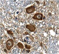
Immunohistochemistry Analysis: A 1:50 dilution from a representative lot detected RA-induced protein 17 in human thalamus tissue.
Biological Information
Immunogen
Recombinant protein corresponding to human RA-induced protein 17.
Clone
5H5.1
Concentration
Please refer to the Certificate of Analysis for the lot-specific concentration.
~115 kDa observed. Uniprot describes 2 isoforms produced by alternative splicing at ~115 kDa and ~107 kDa.
Physicochemical Information
Dimensions
Materials Information
Toxicological Information
Safety Information according to GHS
Safety Information
Product Usage Statements
Quality Assurance
Evaluated by Western Blotting in SH-SY5Y cell lysate.
Western Blotting Analysis: 0.5 µg/mL of this antibody detected RA-induced protein 17 in 10 µg of SH-SY5Y cell lysate.
Usage Statement
Unless otherwise stated in our catalog or other company documentation accompanying the product(s), our products are intended for research use only and are not to be used for any other purpose, which includes but is not limited to, unauthorized commercial uses, in vitro diagnostic uses, ex vivo or in vivo therapeutic uses or any type of consumption or application to humans or animals.
Storage and Shipping Information
Storage Conditions
Stable for 1 year at 2-8°C from date of receipt.
Packaging Information
Material Size
100 µg
Transport Information
Supplemental Information
Specifications
Global Trade Item Number
Bestellnummer
GTIN
MABN250
04053252923913
Documentation
Anti-RA-induced protein 17 Antibody, clone 5H5.1 SDB