Wenn Sie das Fenster schließen, wird Ihre Konfiguration nicht gespeichert, es sei denn, Sie haben Ihren Artikel in die Bestellung aufgenommen oder zu Ihren Favoriten hinzugefügt.
Klicken Sie auf OK, um das MILLIPLEX® MAP-Tool zu schließen oder auf Abbrechen, um zu Ihrer Auswahl zurückzukehren.
Wählen Sie konfigurierbare Panels & Premixed-Kits - ODER - Kits für die zelluläre Signaltransduktion & MAPmates™
Konfigurieren Sie Ihre MILLIPLEX® MAP-Kits und lassen sich den Preis anzeigen.
Konfigurierbare Panels & Premixed-Kits
Unser breites Angebot enthält Multiplex-Panels, für die Sie die Analyten auswählen können, die am besten für Ihre Anwendung geeignet sind. Unter einem separaten Register können Sie das Premixed-Cytokin-Format oder ein Singleplex-Kit wählen.
Kits für die zelluläre Signaltransduktion & MAPmates™
Wählen Sie gebrauchsfertige Kits zur Erforschung gesamter Signalwege oder Prozesse. Oder konfigurieren Sie Ihre eigenen Kits mit Singleplex MAPmates™.
Die folgenden MAPmates™ sollten nicht zusammen analysiert werden: -MAPmates™, die einen unterschiedlichen Assaypuffer erfordern. -Phosphospezifische und MAPmate™ Gesamtkombinationen wie Gesamt-GSK3β und Gesamt-GSK3β (Ser 9). -PanTyr und locusspezifische MAPmates™, z.B. Phospho-EGF-Rezeptor und Phospho-STAT1 (Tyr701). -Mehr als 1 Phospho-MAPmate™ für ein einziges Target (Akt, STAT3). -GAPDH und β-Tubulin können nicht mit Kits oder MAPmates™, die panTyr enthalten, analysiert werden.
.
Bestellnummer
Bestellinformationen
St./Pkg.
Liste
Dieser Artikel wurde zu Ihren Favoriten hinzugefügt.
Wählen Sie bitte Spezies, Panelart, Kit oder Probenart
Um Ihr MILLIPLEX® MAP-Kit zu konfigurieren, wählen Sie zunächst eine Spezies, eine Panelart und/oder ein Kit.
Custom Premix Selecting "Custom Premix" option means that all of the beads you have chosen will be premixed in manufacturing before the kit is sent to you.
Catalogue Number
Ordering Description
Qty/Pack
List
Dieser Artikel wurde zu Ihren Favoriten hinzugefügt.
Spezies
Panelart
Gewähltes Kit
Menge
Bestellnummer
Bestellinformationen
St./Pkg.
Listenpreis
96-Well Plate
Menge
Bestellnummer
Bestellinformationen
St./Pkg.
Listenpreis
Weitere Reagenzien hinzufügen (MAPmates erfordern die Verwendung eines Puffer- und Detektionskits)
Menge
Bestellnummer
Bestellinformationen
St./Pkg.
Listenpreis
48-602MAG
Buffer Detection Kit for Magnetic Beads
1 Kit
Platzsparende Option Kunden, die mehrere Kits kaufen, können ihre Multiplex-Assaykomponenten in Kunststoffbeuteln anstelle von Packungen erhalten, um eine kompaktere Lagerung zu ermöglichen.
Dieser Artikel wurde zu Ihren Favoriten hinzugefügt.
Das Produkt wurde in Ihre Bestellung aufgenommen
Sie können nun ein weiteres Kit konfigurieren, ein Premixed-Kit wählen, zur Kasse gehen oder das Bestell-Tool schließen.
This Anti-Perilipin-3 antibody, C-terminus is validated for use in western blotting & ICC for the detection of Perilipin-3.
More>>This Anti-Perilipin-3 antibody, C-terminus is validated for use in western blotting & ICC for the detection of Perilipin-3. Less<<
Anti-Perilipin-3, C-terminus Antibody: SDB (Sicherheitsdatenblätter), Analysenzertifikate und Qualitätszertifikate, Dossiers, Broschüren und andere verfügbare Dokumente.
Perilipin-3, also known as Cargo selection protein TIP47, or Placental protein 17, PP17, 47 kDa mannose 6-phosphate receptor-binding protein, 47 kDa MPR-binding protein, also known as Mannose-6-phosphate receptor-binding protein 1, and encoded by the gene PLIN3/M6prbp1/Tip47, is a peripheral membrane protein that is required for the transport of mannose 6-phosphate receptors (MPR) from the endosomes to the trans-Golgi network. Perilipin 3 is part of the Perilipin family of proteins which are proteins that decorate the surface of cytosolic lipid droplets and play important roles in lipid metabolism. Cytosolic lipid droplets are now recognized as multifunctional organelles that, among other things, can accumulate in leukocytes under various inflammatory conditions. Perilipin-3 seems to play a critical role in the formation of lipid droplets and production of the the prostaglandin PGE2 an important inflammatory compound.
References
Product Information
Format
Affinity Purified
Presentation
Purified rabbit polyclonal in buffer containing PBS without preservatives with 10% glycerol.
This Anti-Perilipin-3 antibody, C-terminus is validated for use in western blotting & ICC for the detection of Perilipin-3.
Key Applications
Western Blotting
Immunocytochemistry
Application Notes
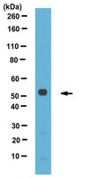
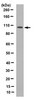
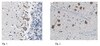
Immunocytochemistry Analysis: A representative lot detected Perilipin-3, C-terminus in OP9 cells (Skinner, J. R., et al. (2009). J Biol Chem. 284(45):30941-30948).
Biological Information
Immunogen
Linear peptide corresponding to the C-terminus of mouse Perilipin-3, C-terminus.
Epitope
C-terminus
Concentration
Please refer to the Certificate of Analysis for the lot-specific concentration.
Evaluated by Western Blotting in NIH/3T3-L1 cell lysate.
Western Blotting Analysis: A 1:1,000 dilution of this antibody detected Perilipin-3, C-terminus in 10 µg of NIH/3T3-L1 cell lysate.
Usage Statement
Unless otherwise stated in our catalog or other company documentation accompanying the product(s), our products are intended for research use only and are not to be used for any other purpose, which includes but is not limited to, unauthorized commercial uses, in vitro diagnostic uses, ex vivo or in vivo therapeutic uses or any type of consumption or application to humans or animals.
Storage and Shipping Information
Storage Conditions
Stable for 1 year at -20°C from date of receipt. Handling Recommendations: Upon receipt and prior to removing the cap, centrifuge the vial and gently mix the solution. Aliquot into microcentrifuge tubes and store at -20°C. Avoid repeated freeze/thaw cycles, which may damage IgG and affect product performance. Note: Variability in freezer temperatures below -20°C may cause glycerol containing solutions to become frozen during storage.
Diacylglycerol enrichment of endoplasmic reticulum or lipid droplets recruits perilipin 3/TIP47 during lipid storage and mobilization. Skinner, James R, et al. J. Biol. Chem., 284: 30941-8 (2009)
2009
Fatty acid-induced triacylglycerol synthesis produces triacylglycerol droplets with a protein coat that includes perilipin 3/TIP47 and perilipin 4/S3-12. This study addresses the following two questions. Where do lipid droplets emerge, and how are their coat proteins recruited? We show that perilipin 3- and perilipin 4-coated lipid droplets emerge along the endoplasmic reticulum (ER). Blocking membrane trafficking with AlF(4)(-) during fatty acid-induced triacylglycerol synthesis drove perilipin 3 to the tubular ER. Forskolin, which like AlF(4)(-) activates adenylate cyclase, did not redistribute perilipin 3, but when added together with AlF(4)(-) perilipin 3 was recruited to lipid droplets rather than the ER. Thus inhibiting trafficking with AlF(4)(-) redistributed perilipin 3 differently under conditions of triacylglycerol synthesis (fatty acid addition) versus hydrolysis (forskolin) suggesting a shared acylglycerol-mediated mechanism. We tested whether diacylglycerol (DG), the immediate precursor of triacylglycerol and its first hydrolytic product, affects the distribution of perilipin 3. Stabilizing DG with the DG lipase inhibitor RHC80267 enhanced the perilipin 3 recruited to lipid droplets and raised DG levels in this fraction. Treating cells with a membrane-permeable DG recruited perilipin 3 to the ER. Stabilizing DG, by blocking its hydrolysis with RHC80267 or its acylation with triacsin C, enhanced recruitment of perilipin 3 to the ER. Expressing the ER enzyme DGAT1, which removes DG by converting it to triacylglycerol, attenuated perilipin 3 DG-induced ER recruitment. Membrane-permeable DG also drove perilipin 4 and 5 onto the ER. Together the data suggest that these lipid droplet proteins are recruited to DG-enriched membranes thereby linking lipid coat proteins to the metabolic state of the cell.