Wenn Sie das Fenster schließen, wird Ihre Konfiguration nicht gespeichert, es sei denn, Sie haben Ihren Artikel in die Bestellung aufgenommen oder zu Ihren Favoriten hinzugefügt.
Klicken Sie auf OK, um das MILLIPLEX® MAP-Tool zu schließen oder auf Abbrechen, um zu Ihrer Auswahl zurückzukehren.
Wählen Sie konfigurierbare Panels & Premixed-Kits - ODER - Kits für die zelluläre Signaltransduktion & MAPmates™
Konfigurieren Sie Ihre MILLIPLEX® MAP-Kits und lassen sich den Preis anzeigen.
Konfigurierbare Panels & Premixed-Kits
Unser breites Angebot enthält Multiplex-Panels, für die Sie die Analyten auswählen können, die am besten für Ihre Anwendung geeignet sind. Unter einem separaten Register können Sie das Premixed-Cytokin-Format oder ein Singleplex-Kit wählen.
Kits für die zelluläre Signaltransduktion & MAPmates™
Wählen Sie gebrauchsfertige Kits zur Erforschung gesamter Signalwege oder Prozesse. Oder konfigurieren Sie Ihre eigenen Kits mit Singleplex MAPmates™.
Die folgenden MAPmates™ sollten nicht zusammen analysiert werden: -MAPmates™, die einen unterschiedlichen Assaypuffer erfordern. -Phosphospezifische und MAPmate™ Gesamtkombinationen wie Gesamt-GSK3β und Gesamt-GSK3β (Ser 9). -PanTyr und locusspezifische MAPmates™, z.B. Phospho-EGF-Rezeptor und Phospho-STAT1 (Tyr701). -Mehr als 1 Phospho-MAPmate™ für ein einziges Target (Akt, STAT3). -GAPDH und β-Tubulin können nicht mit Kits oder MAPmates™, die panTyr enthalten, analysiert werden.
.
Bestellnummer
Bestellinformationen
St./Pkg.
Liste
Dieser Artikel wurde zu Ihren Favoriten hinzugefügt.
Wählen Sie bitte Spezies, Panelart, Kit oder Probenart
Um Ihr MILLIPLEX® MAP-Kit zu konfigurieren, wählen Sie zunächst eine Spezies, eine Panelart und/oder ein Kit.
Custom Premix Selecting "Custom Premix" option means that all of the beads you have chosen will be premixed in manufacturing before the kit is sent to you.
Catalogue Number
Ordering Description
Qty/Pack
List
Dieser Artikel wurde zu Ihren Favoriten hinzugefügt.
Spezies
Panelart
Gewähltes Kit
Menge
Bestellnummer
Bestellinformationen
St./Pkg.
Listenpreis
96-Well Plate
Menge
Bestellnummer
Bestellinformationen
St./Pkg.
Listenpreis
Weitere Reagenzien hinzufügen (MAPmates erfordern die Verwendung eines Puffer- und Detektionskits)
Menge
Bestellnummer
Bestellinformationen
St./Pkg.
Listenpreis
48-602MAG
Buffer Detection Kit for Magnetic Beads
1 Kit
Platzsparende Option Kunden, die mehrere Kits kaufen, können ihre Multiplex-Assaykomponenten in Kunststoffbeuteln anstelle von Packungen erhalten, um eine kompaktere Lagerung zu ermöglichen.
Dieser Artikel wurde zu Ihren Favoriten hinzugefügt.
Das Produkt wurde in Ihre Bestellung aufgenommen
Sie können nun ein weiteres Kit konfigurieren, ein Premixed-Kit wählen, zur Kasse gehen oder das Bestell-Tool schließen.
NE1017
Sigma-AldrichAnti-Pan-Neuronal Neurofilament Marker Mouse mAb (SMI-311)
This Anti-Pan-Neuronal Neurofilament Marker Mouse mAb (SMI-311) is validated for use in ELISA, Frozen Sections, WB, ICC, Paraffin Sections for the detection of Pan-Neuronal Neurofilament Marker.
More>>This Anti-Pan-Neuronal Neurofilament Marker Mouse mAb (SMI-311) is validated for use in ELISA, Frozen Sections, WB, ICC, Paraffin Sections for the detection of Pan-Neuronal Neurofilament Marker. Less<<
SDB (Sicherheitsdatenblätter), Analysenzertifikate und Qualitätszertifikate, Dossiers, Broschüren und andere verfügbare Dokumente.
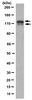
Recognizes ~180-200 kDa pan-neuronal neurofilament marker in rat central nervous system cytoskeletal preparations.
Catalogue Number
NE1017
Brand Family
Calbiochem®
Application Data
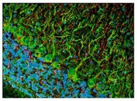
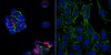
Detection of rat pan-neuronal neurofilament marker by staining frozen sections. Sample: Rat brain. Primary antibody: Anti-Pan-Neuronal Neurofilament Marker Mouse mAb (SMI-311) (Cat. No. NE1017) (1:1000). Detection: fluorescence (green) with Hoechst 33342 counterstain.
References
References
Agostino, A., et al. 2003. Hum. Mol. Genet.12, 399. Tsunoda, I., et al. 2003. Am. J. Pathol.162, 1259. Ulfig, N., et al. 1998. Cell Tissue Res.291, 433.
ELISA (1:1000) Frozen Sections (1:1000, see comments) Immunoblotting (1:1000) Immunocytochemistry (1:1000, see comments) Paraffin Sections (1:1000, heat pre-treatment required, see comments)
Application Comments
This antibody cocktail was selected to provide a specific marker for neurons in tissue sections and cultured cells. In contrast to individual anti-nonphosphoneurofilament antibodies that identify different subsets of neurons, this cocktail is a convenient marker for neurons in general and their differentiation from non-neuronal cells. Also useful as an early marker of neuronal migration and differentiation in human fetal development, yielding Golgi-like images without the disadvantages of the lack of selectivity and poor specificity of the Golgi technique. Can be used to trace the "inside-out gradient" of neuron production and differentiation in specifically delineating cell bodies and dendrites. Certain pathologic conditions, such as malnutrition, affect the SMI-311-visualized soma size and dendritic arborization. Tissues and cultured cells can be fixed in a variety of paraformaldehyde- or formaldehyde-containing fixatives, including Bouin's fixative. This antibody does not react well with tissues or cells fixed in glutaraldehyde/paraformaldehyde. For staining paraffin sections it is recommended that de-paraffinized tissues be autoclaved in dH2O for 10 min to expose the epitope. Trypsin pre-treatment will abolish reactivity. Post-fixation in cold methanol or methanol/hydrogen peroxide facilitates access of the antibody to the neurons in frozen sections and thick tissues sections that have been fixed in 4% paraformaldehyde or cultured cells. For tissues that have not been paraffin-embedded and have been stored for long periods of time in formaldehyde fixatives, it is recommended that the tissues (up to 0.5 cm thick) be boiled in Tris-buffered saline, pH 9.0 for 15 min prior to sectioning. Antibody should be titrated for optimal results in individual systems.
Biological Information
Immunogen
homogenized hypothalami extracted from Fischer 344 rat brain
Immunogen
Rat
Clone
SMI-311
Host
Mouse
Isotype
IgG₁, IgM cocktail
Species Reactivity
Mammals
Antibody Type
Monoclonal Antibody
Physicochemical Information
Dimensions
Materials Information
Toxicological Information
Safety Information according to GHS
Safety Information
Product Usage Statements
Storage and Shipping Information
Ship Code
Dry Ice Only
Toxicity
Standard Handling
Storage
-20°C
Avoid freeze/thaw
Avoid freeze/thaw
Do not freeze
Ok to freeze
Special Instructions
Following initial thaw, aliquot and freeze (-20°C).
Packaging Information
Transport Information
Supplemental Information
Specifications
Global Trade Item Number
Bestellnummer
GTIN
NE1017
0
Documentation
Anti-Pan-Neuronal Neurofilament Marker Mouse mAb (SMI-311) SDB
Anti-Pan-Neuronal Neurofilament Marker Mouse mAb (SMI-311) Analysenzertifikate
Titel
Chargennummer
NE1017
Literatur
Übersicht
Agostino, A., et al. 2003. Hum. Mol. Genet.12, 399. Tsunoda, I., et al. 2003. Am. J. Pathol.162, 1259. Ulfig, N., et al. 1998. Cell Tissue Res.291, 433.
Datenblatt
Note that this data sheet is not lot-specific and is representative of the current specifications for this product. Please consult the vial label and the certificate of analysis for information on specific lots. Also note that shipping conditions may differ from storage conditions.
Revision
01-October-2007 RFH
Application
ELISA (1:1000) Frozen Sections (1:1000, see comments) Immunoblotting (1:1000) Immunocytochemistry (1:1000, see comments) Paraffin Sections (1:1000, heat pre-treatment required, see comments)
Application Data
Detection of rat pan-neuronal neurofilament marker by staining frozen sections. Sample: Rat brain. Primary antibody: Anti-Pan-Neuronal Neurofilament Marker Mouse mAb (SMI-311) (Cat. No. NE1017) (1:1000). Detection: fluorescence (green) with Hoechst 33342 counterstain.
Description
Mouse monoclonal antibody supplied as undiluted ascites. Recognizes the ~180-200 kDa pan-neuronal neurofilament marker protein.
Background
Pan-neuronal neurofilament marker is a specific marker for neurons.
Host
Mouse
Immunogen species
Rat
Immunogen
homogenized hypothalami extracted from Fischer 344 rat brain
Clone
SMI-311
Isotype
IgG₁, IgM cocktail
Species
mammalian
Positive control
Rat brain or rat CNS cytoskeletal preparations
Form
Liquid
Formulation
Undiluted ascites.
Preservative
≤0.1% sodium azide
Comments
This antibody cocktail was selected to provide a specific marker for neurons in tissue sections and cultured cells. In contrast to individual anti-nonphosphoneurofilament antibodies that identify different subsets of neurons, this cocktail is a convenient marker for neurons in general and their differentiation from non-neuronal cells. Also useful as an early marker of neuronal migration and differentiation in human fetal development, yielding Golgi-like images without the disadvantages of the lack of selectivity and poor specificity of the Golgi technique. Can be used to trace the "inside-out gradient" of neuron production and differentiation in specifically delineating cell bodies and dendrites. Certain pathologic conditions, such as malnutrition, affect the SMI-311-visualized soma size and dendritic arborization. Tissues and cultured cells can be fixed in a variety of paraformaldehyde- or formaldehyde-containing fixatives, including Bouin's fixative. This antibody does not react well with tissues or cells fixed in glutaraldehyde/paraformaldehyde. For staining paraffin sections it is recommended that de-paraffinized tissues be autoclaved in dH2O for 10 min to expose the epitope. Trypsin pre-treatment will abolish reactivity. Post-fixation in cold methanol or methanol/hydrogen peroxide facilitates access of the antibody to the neurons in frozen sections and thick tissues sections that have been fixed in 4% paraformaldehyde or cultured cells. For tissues that have not been paraffin-embedded and have been stored for long periods of time in formaldehyde fixatives, it is recommended that the tissues (up to 0.5 cm thick) be boiled in Tris-buffered saline, pH 9.0 for 15 min prior to sectioning. Antibody should be titrated for optimal results in individual systems.
Storage
Avoid freeze/thaw
-20°C
Do Not Freeze
Ok to freeze
Special Instructions
Following initial thaw, aliquot and freeze (-20°C).
Toxicity
Standard Handling
References
Agostino, A., et al. 2003. Hum. Mol. Genet.12, 399. Tsunoda, I., et al. 2003. Am. J. Pathol.162, 1259. Ulfig, N., et al. 1998. Cell Tissue Res.291, 433.