Wenn Sie das Fenster schließen, wird Ihre Konfiguration nicht gespeichert, es sei denn, Sie haben Ihren Artikel in die Bestellung aufgenommen oder zu Ihren Favoriten hinzugefügt.
Klicken Sie auf OK, um das MILLIPLEX® MAP-Tool zu schließen oder auf Abbrechen, um zu Ihrer Auswahl zurückzukehren.
Wählen Sie konfigurierbare Panels & Premixed-Kits - ODER - Kits für die zelluläre Signaltransduktion & MAPmates™
Konfigurieren Sie Ihre MILLIPLEX® MAP-Kits und lassen sich den Preis anzeigen.
Konfigurierbare Panels & Premixed-Kits
Unser breites Angebot enthält Multiplex-Panels, für die Sie die Analyten auswählen können, die am besten für Ihre Anwendung geeignet sind. Unter einem separaten Register können Sie das Premixed-Cytokin-Format oder ein Singleplex-Kit wählen.
Kits für die zelluläre Signaltransduktion & MAPmates™
Wählen Sie gebrauchsfertige Kits zur Erforschung gesamter Signalwege oder Prozesse. Oder konfigurieren Sie Ihre eigenen Kits mit Singleplex MAPmates™.
Die folgenden MAPmates™ sollten nicht zusammen analysiert werden: -MAPmates™, die einen unterschiedlichen Assaypuffer erfordern. -Phosphospezifische und MAPmate™ Gesamtkombinationen wie Gesamt-GSK3β und Gesamt-GSK3β (Ser 9). -PanTyr und locusspezifische MAPmates™, z.B. Phospho-EGF-Rezeptor und Phospho-STAT1 (Tyr701). -Mehr als 1 Phospho-MAPmate™ für ein einziges Target (Akt, STAT3). -GAPDH und β-Tubulin können nicht mit Kits oder MAPmates™, die panTyr enthalten, analysiert werden.
.
Bestellnummer
Bestellinformationen
St./Pkg.
Liste
Dieser Artikel wurde zu Ihren Favoriten hinzugefügt.
Wählen Sie bitte Spezies, Panelart, Kit oder Probenart
Um Ihr MILLIPLEX® MAP-Kit zu konfigurieren, wählen Sie zunächst eine Spezies, eine Panelart und/oder ein Kit.
Custom Premix Selecting "Custom Premix" option means that all of the beads you have chosen will be premixed in manufacturing before the kit is sent to you.
Catalogue Number
Ordering Description
Qty/Pack
List
Dieser Artikel wurde zu Ihren Favoriten hinzugefügt.
Spezies
Panelart
Gewähltes Kit
Menge
Bestellnummer
Bestellinformationen
St./Pkg.
Listenpreis
96-Well Plate
Menge
Bestellnummer
Bestellinformationen
St./Pkg.
Listenpreis
Weitere Reagenzien hinzufügen (MAPmates erfordern die Verwendung eines Puffer- und Detektionskits)
Menge
Bestellnummer
Bestellinformationen
St./Pkg.
Listenpreis
48-602MAG
Buffer Detection Kit for Magnetic Beads
1 Kit
Platzsparende Option Kunden, die mehrere Kits kaufen, können ihre Multiplex-Assaykomponenten in Kunststoffbeuteln anstelle von Packungen erhalten, um eine kompaktere Lagerung zu ermöglichen.
Dieser Artikel wurde zu Ihren Favoriten hinzugefügt.
Das Produkt wurde in Ihre Bestellung aufgenommen
Sie können nun ein weiteres Kit konfigurieren, ein Premixed-Kit wählen, zur Kasse gehen oder das Bestell-Tool schließen.
CA1029
Sigma-AldrichAnti-N-Cadherin Mouse mAb (13A9)
Anti-N-Cadherin Mouse mAb (13A9): SDB (Sicherheitsdatenblätter), Analysenzertifikate und Qualitätszertifikate, Dossiers, Broschüren und andere verfügbare Dokumente.
Recognizes the ~140 kDa N-cadherin protein in HeLa cells.
Catalogue Number
CA1029
Brand Family
Calbiochem®
Application Data
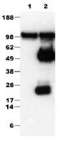
Lane 1: Detection of human N-cadherin by immunoblotting. Sample: Whole cell lysate (25 µg) from HeLa cells. Primary antibody: Anti-N-Cadherin Mouse mAb (13A9) (Cat. No. CA1029) (1:1000). Detection: chemiluminescence.
Lane 2: Detection of human N-cadherin by immunoprecipitation followed by immunoblotting. Antibody for immunoprecipitation: Anti N-Cadherin Mouse mAb (13A9) (Cat No. CA1029) (1:100/500 µg total protein). Immunoblotting conditions: same as lane 1.
References
References
Christofori, G. 2006. Nature44, 444. Cvallaro, U., et al. 2006. Exp. Cell Res.312, 659. Cowin, P., et al. 2005. Curr. Op. Cell Biol.17, 499.
Antibody should be titrated for optimal results in individual systems.
Biological Information
Immunogen
a recombinant fusion protein consisting of the cytoplasmic domain of human N-cadherin, expressed in E. coli
Immunogen
Human
Clone
13A9
Host
Mouse
Isotype
IgG
Physicochemical Information
Dimensions
Materials Information
Toxicological Information
Safety Information according to GHS
Safety Information
Product Usage Statements
Storage and Shipping Information
Ship Code
Blue Ice Only
Toxicity
Standard Handling
Storage
-20°C
Avoid freeze/thaw
Avoid freeze/thaw
Do not freeze
Ok to freeze
Special Instructions
Following initial thaw, aliquot and freeze (-20°C).
Packaging Information
Transport Information
Supplemental Information
Specifications
Global Trade Item Number
Bestellnummer
GTIN
CA1029
0
Documentation
Literatur
Übersicht
Christofori, G. 2006. Nature44, 444. Cvallaro, U., et al. 2006. Exp. Cell Res.312, 659. Cowin, P., et al. 2005. Curr. Op. Cell Biol.17, 499.
Datenblatt
Note that this data sheet is not lot-specific and is representative of the current specifications for this product. Please consult the vial label and the certificate of analysis for information on specific lots. Also note that shipping conditions may differ from storage conditions.
Lane 1: Detection of human N-cadherin by immunoblotting. Sample: Whole cell lysate (25 µg) from HeLa cells. Primary antibody: Anti-N-Cadherin Mouse mAb (13A9) (Cat. No. CA1029) (1:1000). Detection: chemiluminescence.
Lane 2: Detection of human N-cadherin by immunoprecipitation followed by immunoblotting. Antibody for immunoprecipitation: Anti N-Cadherin Mouse mAb (13A9) (Cat No. CA1029) (1:100/500 µg total protein). Immunoblotting conditions: same as lane 1.
Description
Mouse monoclonal antibody supplied as undiluted ascites. Recognizes the ~140 kDa N-cadherin protein.
Background
Cadherins are a large family of Ca2+-dependant cell adhesion transmembrane glycoproteins that play a role in cell to cell recognition. The classic cadherins include N-, R-, P-, B-, and E-cadherin. Although cadherins interact with similar intracellular proteins, they can exert specifc activities. Early non-invasive epithelial cells express E-cadherin. However, during the the process of epithelial to mesenchymal transition (EMT), the expression of E-cadherin is lost and the expression of N-cadherin is upregulated. This process, the cadherin switch, results in the transition of a benign tumor to a malignant and metastatic form. N-cadherin is a 140-kDa protein that has several functions that contribute to tumor invasion. It promotes cell-cell adhesion to N-cadherin stromal cells and it binds to and activates FGFRs that result in activation of the PI 3-K and MAPK pathways. N-cadherin is also cleaved by MMPs and a γ-secretase-like protease, releasing the extracellular domain of N-cadherin and the intracellular carboxy terminal.
Host
Mouse
Immunogen species
Human
Immunogen
a recombinant fusion protein consisting of the cytoplasmic domain of human N-cadherin, expressed in E. coli
Clone
13A9
Isotype
IgG
Species
human
Positive control
HeLa cells
Form
Liquid
Formulation
Undiluted ascites.
Preservative
≤0.1% sodium azide
Comments
Antibody should be titrated for optimal results in individual systems.
Storage
Avoid freeze/thaw
-20°C
Do Not Freeze
Ok to freeze
Special Instructions
Following initial thaw, aliquot and freeze (-20°C).
Toxicity
Standard Handling
References
Christofori, G. 2006. Nature44, 444. Cvallaro, U., et al. 2006. Exp. Cell Res.312, 659. Cowin, P., et al. 2005. Curr. Op. Cell Biol.17, 499.