Wenn Sie das Fenster schließen, wird Ihre Konfiguration nicht gespeichert, es sei denn, Sie haben Ihren Artikel in die Bestellung aufgenommen oder zu Ihren Favoriten hinzugefügt.
Klicken Sie auf OK, um das MILLIPLEX® MAP-Tool zu schließen oder auf Abbrechen, um zu Ihrer Auswahl zurückzukehren.
Wählen Sie konfigurierbare Panels & Premixed-Kits - ODER - Kits für die zelluläre Signaltransduktion & MAPmates™
Konfigurieren Sie Ihre MILLIPLEX® MAP-Kits und lassen sich den Preis anzeigen.
Konfigurierbare Panels & Premixed-Kits
Unser breites Angebot enthält Multiplex-Panels, für die Sie die Analyten auswählen können, die am besten für Ihre Anwendung geeignet sind. Unter einem separaten Register können Sie das Premixed-Cytokin-Format oder ein Singleplex-Kit wählen.
Kits für die zelluläre Signaltransduktion & MAPmates™
Wählen Sie gebrauchsfertige Kits zur Erforschung gesamter Signalwege oder Prozesse. Oder konfigurieren Sie Ihre eigenen Kits mit Singleplex MAPmates™.
Die folgenden MAPmates™ sollten nicht zusammen analysiert werden: -MAPmates™, die einen unterschiedlichen Assaypuffer erfordern. -Phosphospezifische und MAPmate™ Gesamtkombinationen wie Gesamt-GSK3β und Gesamt-GSK3β (Ser 9). -PanTyr und locusspezifische MAPmates™, z.B. Phospho-EGF-Rezeptor und Phospho-STAT1 (Tyr701). -Mehr als 1 Phospho-MAPmate™ für ein einziges Target (Akt, STAT3). -GAPDH und β-Tubulin können nicht mit Kits oder MAPmates™, die panTyr enthalten, analysiert werden.
.
Bestellnummer
Bestellinformationen
St./Pkg.
Liste
Dieser Artikel wurde zu Ihren Favoriten hinzugefügt.
Wählen Sie bitte Spezies, Panelart, Kit oder Probenart
Um Ihr MILLIPLEX® MAP-Kit zu konfigurieren, wählen Sie zunächst eine Spezies, eine Panelart und/oder ein Kit.
Custom Premix Selecting "Custom Premix" option means that all of the beads you have chosen will be premixed in manufacturing before the kit is sent to you.
Catalogue Number
Ordering Description
Qty/Pack
List
Dieser Artikel wurde zu Ihren Favoriten hinzugefügt.
Spezies
Panelart
Gewähltes Kit
Menge
Bestellnummer
Bestellinformationen
St./Pkg.
Listenpreis
96-Well Plate
Menge
Bestellnummer
Bestellinformationen
St./Pkg.
Listenpreis
Weitere Reagenzien hinzufügen (MAPmates erfordern die Verwendung eines Puffer- und Detektionskits)
Menge
Bestellnummer
Bestellinformationen
St./Pkg.
Listenpreis
48-602MAG
Buffer Detection Kit for Magnetic Beads
1 Kit
Platzsparende Option Kunden, die mehrere Kits kaufen, können ihre Multiplex-Assaykomponenten in Kunststoffbeuteln anstelle von Packungen erhalten, um eine kompaktere Lagerung zu ermöglichen.
Dieser Artikel wurde zu Ihren Favoriten hinzugefügt.
Das Produkt wurde in Ihre Bestellung aufgenommen
Sie können nun ein weiteres Kit konfigurieren, ein Premixed-Kit wählen, zur Kasse gehen oder das Bestell-Tool schließen.
This mouse monoclonal Anti-Myosin-3 (MYH3) Antibody, clone BF-G6, Cat. No. MABT818 is validated for use in and Western Blotting, Immunocytochemistry, Immunohistochemistry, Immunofluorescence, ELISA and Western Blotting, for the detection of Myosin-3.
More>>This mouse monoclonal Anti-Myosin-3 (MYH3) Antibody, clone BF-G6, Cat. No. MABT818 is validated for use in and Western Blotting, Immunocytochemistry, Immunohistochemistry, Immunofluorescence, ELISA and Western Blotting, for the detection of Myosin-3. Less<<
Anti-Myosin-3 (MYH3) Antibody, clone BF-G6 : SDB (Sicherheitsdatenblätter), Analysenzertifikate und Qualitätszertifikate, Dossiers, Broschüren und andere verfügbare Dokumente.
Myosin-3 (UniProt A6QPA6; also known as Muscle embryonic myosin heavy chain, Myosin heavy chain 3, MYH3, Protein, Myosin heavy chain, fast skeletal muscle, embryonic, SMHCE) is encoded by the MYH3 gene (Gene ID 281338) in bovine species. Myosin is a major contractile protein that converts chemical energy into mechanical energy through the hydrolysis of ATP. MYH3 is predominantly expressed during fetal development and its expression declines rapidly after birth. It is not normally present in adult muscle except during muscle regeneration. Mutations in MYH3 are known cause two congenital contracture syndromes, Freeman-Sheldon syndrome and Sheldon-Hall syndrome, the most severe forms of distal arthrogryposis (DA). Ref.: Pokrzywa, M., et al. (2015). PLoS One. 10(11):e0142094; Toydamir, R.M., et al. (2006). Nature Genet. 38(5):561-565.
References
Product Information
Format
Purified
Presentation
Purified mouse IgG2b, kappa in buffer containing 0.1 M Tris-Glycine (pH 7.4), 150 mM NaCl with 0.05% sodium azide.
This mouse monoclonal Anti-Myosin-3 (MYH3) Antibody, clone BF-G6, Cat. No. MABT818 is validated for use in and Western Blotting, Immunocytochemistry, Immunohistochemistry, Immunofluorescence, ELISA and Western Blotting, for the detection of Myosin-3.
Key Applications
Western Blotting
Immunohistochemistry
Immunocytochemistry
Immunofluorescence
ELISA
Application Notes
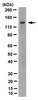
Western Blotting Analysis: 0.5 µg/mL from a representative lot detected Myosin-3 (MYH3) in 10 µg of rat embryonic muscle tissue lysate (Courtesy of Alberto Rossi, Ph.D., University of Colorado, U.S.A.).
ELISA Analysis: Clone BF-G6 hybridoma culture supernatant detected myosin from 10-week human fetus, but not myosin from 8-day new born muscle or adult skeletal muscle (Schiaffino, S., et al. (1986). Exp. Cell Res. 163(1):211-220).
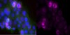
Immunocytochemistry Analysis: Clone BF-G6 hybridoma culture supernatant immunostained aceton-fixed tumor cells in bone marrow aspiration from a child with rhobdomyosarcoma (Schiaffino, S., et al. (1986). Exp. Cell Res. 163(1):211-220).
Immunofluorescence Analysis: Clone BF-G6 hybridoma culture supernatant immunostained serial transverse cryosections of rat extraocular (EO) and soleus muscles. BF-G6 staining pattern is similar, but not identical to that of MYH15 (Rossi, A.C., et al. (2010). J. Physiol. 588(Pt 2):353-364).
Immunofluorescence Analysis: Clone BF-G6 hybridoma culture supernatant immunostained frozen fetal muscle sections, while very few cells were stained in 8-week new born muscle tissue and no staining of 8-month infant muscle tissue was observed (Schiaffino, S., et al. (1986). Exp. Cell Res. 163(1):211-220).
Immunohistochemistry Analysis: Clone BF-G6 hybridoma culture supernatant immunostained tumor cells in frozen human rhobdomyosarcoma sections (Schiaffino, S., et al. (1986). Exp. Cell Res. 163(1):211-220).
Western Blotting Analysis: Clone BF-G6 hybridoma culture supernatant detected myosin from 10-week human fetus, but not myosin from adult skeletal muscle (Schiaffino, S., et al. (1986). Exp. Cell Res. 163(1):211-220).
Clone BF-G6 detected myosin in human fetal muscle, but not adult human skeletal muscle tissue (Schiaffino, S., et al. (1986). Exp. Cell Res. 163(1):211-220).
Isotype
IgG2bκ
Species Reactivity
Human
Rat
Species Reactivity Note
Human, Rat. Predicted to react with Bovine based on 100% sequence homology.
~225 kDa observed. 223.7 kDa (bovine) and 223.9 kDa (human/rat) calculated. Uncharacterized bands may be observed in some lysate(s).
Physicochemical Information
Dimensions
Materials Information
Toxicological Information
Safety Information according to GHS
Safety Information
Product Usage Statements
Quality Assurance
Identity Confirmation by Isotyping Test.
Isotyping Analysis: The identity of this monoclonal antibody is confirmed by isotyping test to be mouse IgG2b, kappa.
Usage Statement
Unless otherwise stated in our catalog or other company documentation accompanying the product(s), our products are intended for research use only and are not to be used for any other purpose, which includes but is not limited to, unauthorized commercial uses, in vitro diagnostic uses, ex vivo or in vivo therapeutic uses or any type of consumption or application to humans or animals.