Wenn Sie das Fenster schließen, wird Ihre Konfiguration nicht gespeichert, es sei denn, Sie haben Ihren Artikel in die Bestellung aufgenommen oder zu Ihren Favoriten hinzugefügt.
Klicken Sie auf OK, um das MILLIPLEX® MAP-Tool zu schließen oder auf Abbrechen, um zu Ihrer Auswahl zurückzukehren.
Wählen Sie konfigurierbare Panels & Premixed-Kits - ODER - Kits für die zelluläre Signaltransduktion & MAPmates™
Konfigurieren Sie Ihre MILLIPLEX® MAP-Kits und lassen sich den Preis anzeigen.
Konfigurierbare Panels & Premixed-Kits
Unser breites Angebot enthält Multiplex-Panels, für die Sie die Analyten auswählen können, die am besten für Ihre Anwendung geeignet sind. Unter einem separaten Register können Sie das Premixed-Cytokin-Format oder ein Singleplex-Kit wählen.
Kits für die zelluläre Signaltransduktion & MAPmates™
Wählen Sie gebrauchsfertige Kits zur Erforschung gesamter Signalwege oder Prozesse. Oder konfigurieren Sie Ihre eigenen Kits mit Singleplex MAPmates™.
Die folgenden MAPmates™ sollten nicht zusammen analysiert werden: -MAPmates™, die einen unterschiedlichen Assaypuffer erfordern. -Phosphospezifische und MAPmate™ Gesamtkombinationen wie Gesamt-GSK3β und Gesamt-GSK3β (Ser 9). -PanTyr und locusspezifische MAPmates™, z.B. Phospho-EGF-Rezeptor und Phospho-STAT1 (Tyr701). -Mehr als 1 Phospho-MAPmate™ für ein einziges Target (Akt, STAT3). -GAPDH und β-Tubulin können nicht mit Kits oder MAPmates™, die panTyr enthalten, analysiert werden.
.
Bestellnummer
Bestellinformationen
St./Pkg.
Liste
Dieser Artikel wurde zu Ihren Favoriten hinzugefügt.
Wählen Sie bitte Spezies, Panelart, Kit oder Probenart
Um Ihr MILLIPLEX® MAP-Kit zu konfigurieren, wählen Sie zunächst eine Spezies, eine Panelart und/oder ein Kit.
Custom Premix Selecting "Custom Premix" option means that all of the beads you have chosen will be premixed in manufacturing before the kit is sent to you.
Catalogue Number
Ordering Description
Qty/Pack
List
Dieser Artikel wurde zu Ihren Favoriten hinzugefügt.
Spezies
Panelart
Gewähltes Kit
Menge
Bestellnummer
Bestellinformationen
St./Pkg.
Listenpreis
96-Well Plate
Menge
Bestellnummer
Bestellinformationen
St./Pkg.
Listenpreis
Weitere Reagenzien hinzufügen (MAPmates erfordern die Verwendung eines Puffer- und Detektionskits)
Menge
Bestellnummer
Bestellinformationen
St./Pkg.
Listenpreis
48-602MAG
Buffer Detection Kit for Magnetic Beads
1 Kit
Platzsparende Option Kunden, die mehrere Kits kaufen, können ihre Multiplex-Assaykomponenten in Kunststoffbeuteln anstelle von Packungen erhalten, um eine kompaktere Lagerung zu ermöglichen.
Dieser Artikel wurde zu Ihren Favoriten hinzugefügt.
Das Produkt wurde in Ihre Bestellung aufgenommen
Sie können nun ein weiteres Kit konfigurieren, ein Premixed-Kit wählen, zur Kasse gehen oder das Bestell-Tool schließen.
Anti-LAMC2, clone P2H, Cat. No. MABT1460, is a mouse monoclonal antibody that detects Laminin subunit g2 and is tested for use in ELISA, Immunocytochemistry, Immunohistochemistry (Paraffin), and Western Blotting.
More>>Anti-LAMC2, clone P2H, Cat. No. MABT1460, is a mouse monoclonal antibody that detects Laminin subunit g2 and is tested for use in ELISA, Immunocytochemistry, Immunohistochemistry (Paraffin), and Western Blotting. Less<<
Laminin subunit g2 (UniProt: Q13753; also known as Cell-scattering factor 140 kDa subunit, CSF 140 kDa subunit, Epiligrin subunit gamma, Kalinin subunit gamma, Kalinin/nicein/epiligrin 100 kDa subunit, Ladsin 140 kDa subunit, Laminin B2t chain, Laminin-5 subunit gamma, Large adhesive scatter factor 140 kDa subunit, Nicein subunit gamma) is encoded by the LAMC2 (also known as LAMB2T, LAMNB2) gene (Gene ID: 3918) in human. Laminin is a major basement membrane glycoprotein consisting of α, β, γ chains that are bound to each other by disulfide bonds. Over 15 different isoforms have been reported in the laminin family that are expressed in a tissue-specific manner and play different role in each tissue. γ² a subunit of Laminin-5 is reported to be frequently over-expressed as a monomer or as the β³-γ² heterodimer in invasive cancers and is regarded as an invasion marker. It has 8 EGF-like domains. The cell adhesion and motility activities of Laminin-5 are reported to be regulated by the proteolytic cleavage of the short arm of Laminin γ² chain by BMP-1 or MT1-MMP. This cleavage releases a 45 kDa N-terminal proteolytic fragment (γ²pf). The short arm and γ²pf can be further cleaved by serine proteases and by matrix metalloproteinases resulting in the release of small N-terminal fragments. γ²pf‐like fragments have been reported to be present in conditioned media of cultured cancer cells and in sera from human cancer subjects. Clone P2H targets the second N-terminal EGF-like repeat (NE2) of domain V of laminin 2. (Ref.: Sato, H., et al. (2014). Cancer Sci. 105(2); 168-175; Miyazaki, K., et al. (2016). Cancer Sci. 107(12); 1909-1918).
References
Product Information
Format
Purified
Presentation
Purified mouse monoclonal antibody IgG2a in buffer containing 0.1 M Tris-Glycine (pH 7.4), 150 mM NaCl with 0.05% sodium azide.
Anti-LAMC2, clone P2H, Cat. No. MABT1460, is a mouse monoclonal antibody that detects Laminin subunit g2 and is tested for use in ELISA, Immunocytochemistry, Immunohistochemistry (Paraffin), and Western Blotting.
Key Applications
Immunocytochemistry
ELISA
Western Blotting
Immunohistochemistry (Paraffin)
Application Notes
Western Blotting Analysis: 1 µg/mL from a representative lot detected LAMC2 in lung squamous cell carcinoma cell lysates (Courtesy of Kaoru Miyazaki, Ph.D., KIhara Institute for Biological Research, Yokohama City University, Japan).
Western Blotting Analysis: A representative lot detected LAMC2 in Western Blotting applications (Miyazaki, K., et. al. (2016). Cancer Sci. 107(12):1909-1918).
Immunohistochemistry (Paraffin) Analysis: 10 µg/mL from a representative lot detected LAMC2 in normal human skin, skin cancer, normal breast, and breast adenocarcinoma tissues (Courtesy of Kaoru Miyazaki, Ph.D., KIhara Institute for Biological Research, Yokohama City University, Japan).
ELISA Analysis: 5 µg/mL from a representative lot detected LAMC2 in Lm-gamma2pf and Lm-gamma2 in Lm332 condition medium from Lm332-expressing HEK293 cells (Courtesy of Kaoru Miyazaki, Ph.D., KIhara Institute for Biological Research, Yokohama City University, Japan).
Immunocytochemistry Analysis: A representative lot detected LAMC2 in Immunocytochemistry applications (Miyazaki, K., et. al. (2016). Cancer Sci. 107(12):1909-1918).
Immunohistochemistry Applications: A representative lot detected LAMC2 in Immunohistochemistry applications of lung squamous cell carcinomas and lung adenocarcinomas (Courtesy of Kaoru Miyazaki, Ph.D., KIhara Institute for Biological Research, Yokohama City University, Japan).
Note: Actual optimal working dilutions must be determined by end user as specimens, and experimental conditions may vary with the end user
Biological Information
Immunogen
His-tagged 45-kDa N-terminal fragment consisting of g2pf, domain V.
Epitope
N-terminal
Clone
P2H
Concentration
0.5 mg/mL. Please refer to guidance on suggested starting dilutions and/or titers per application and sample type.
Host
Mouse
Specificity
Clone P2H is a mouse monoclonal antibody that detects Laminin-5, g2 chain. It targets an epitope within the N-terminal, NE2 in domain V.
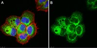
Immunocytochemistry Analysis: A 1:500 dilution of this antibody detected Laminin-5 subunit g2 (LAMC2) in A431 cells.
Usage Statement
Unless otherwise stated in our catalog or other company documentation accompanying the product(s), our products are intended for research use only and are not to be used for any other purpose, which includes but is not limited to, unauthorized commercial uses, in vitro diagnostic uses, ex vivo or in vivo therapeutic uses or any type of consumption or application to humans or animals.
Storage and Shipping Information
Storage Conditions
Stable for 1 year at +2°C to +8°C from date of receipt.