Wenn Sie das Fenster schließen, wird Ihre Konfiguration nicht gespeichert, es sei denn, Sie haben Ihren Artikel in die Bestellung aufgenommen oder zu Ihren Favoriten hinzugefügt.
Klicken Sie auf OK, um das MILLIPLEX® MAP-Tool zu schließen oder auf Abbrechen, um zu Ihrer Auswahl zurückzukehren.
Wählen Sie konfigurierbare Panels & Premixed-Kits - ODER - Kits für die zelluläre Signaltransduktion & MAPmates™
Konfigurieren Sie Ihre MILLIPLEX® MAP-Kits und lassen sich den Preis anzeigen.
Konfigurierbare Panels & Premixed-Kits
Unser breites Angebot enthält Multiplex-Panels, für die Sie die Analyten auswählen können, die am besten für Ihre Anwendung geeignet sind. Unter einem separaten Register können Sie das Premixed-Cytokin-Format oder ein Singleplex-Kit wählen.
Kits für die zelluläre Signaltransduktion & MAPmates™
Wählen Sie gebrauchsfertige Kits zur Erforschung gesamter Signalwege oder Prozesse. Oder konfigurieren Sie Ihre eigenen Kits mit Singleplex MAPmates™.
Die folgenden MAPmates™ sollten nicht zusammen analysiert werden: -MAPmates™, die einen unterschiedlichen Assaypuffer erfordern. -Phosphospezifische und MAPmate™ Gesamtkombinationen wie Gesamt-GSK3β und Gesamt-GSK3β (Ser 9). -PanTyr und locusspezifische MAPmates™, z.B. Phospho-EGF-Rezeptor und Phospho-STAT1 (Tyr701). -Mehr als 1 Phospho-MAPmate™ für ein einziges Target (Akt, STAT3). -GAPDH und β-Tubulin können nicht mit Kits oder MAPmates™, die panTyr enthalten, analysiert werden.
.
Bestellnummer
Bestellinformationen
St./Pkg.
Liste
Dieser Artikel wurde zu Ihren Favoriten hinzugefügt.
Wählen Sie bitte Spezies, Panelart, Kit oder Probenart
Um Ihr MILLIPLEX® MAP-Kit zu konfigurieren, wählen Sie zunächst eine Spezies, eine Panelart und/oder ein Kit.
Custom Premix Selecting "Custom Premix" option means that all of the beads you have chosen will be premixed in manufacturing before the kit is sent to you.
Catalogue Number
Ordering Description
Qty/Pack
List
Dieser Artikel wurde zu Ihren Favoriten hinzugefügt.
Spezies
Panelart
Gewähltes Kit
Menge
Bestellnummer
Bestellinformationen
St./Pkg.
Listenpreis
96-Well Plate
Menge
Bestellnummer
Bestellinformationen
St./Pkg.
Listenpreis
Weitere Reagenzien hinzufügen (MAPmates erfordern die Verwendung eines Puffer- und Detektionskits)
Menge
Bestellnummer
Bestellinformationen
St./Pkg.
Listenpreis
48-602MAG
Buffer Detection Kit for Magnetic Beads
1 Kit
Platzsparende Option Kunden, die mehrere Kits kaufen, können ihre Multiplex-Assaykomponenten in Kunststoffbeuteln anstelle von Packungen erhalten, um eine kompaktere Lagerung zu ermöglichen.
Dieser Artikel wurde zu Ihren Favoriten hinzugefügt.
Das Produkt wurde in Ihre Bestellung aufgenommen
Sie können nun ein weiteres Kit konfigurieren, ein Premixed-Kit wählen, zur Kasse gehen oder das Bestell-Tool schließen.
Use Anti-IL-7Ra (CD127) Antibody, extracellular (Rabbit Polyclonal Antibody) validated in ELISA, WB, FC to detect IL-7Ra (CD127) also known as CD127 antigen, interleukin 7 receptor.
More>>Use Anti-IL-7Ra (CD127) Antibody, extracellular (Rabbit Polyclonal Antibody) validated in ELISA, WB, FC to detect IL-7Ra (CD127) also known as CD127 antigen, interleukin 7 receptor. Less<<
Anti-IL-7Ra (CD127) Antibody, extracellular: SDB (Sicherheitsdatenblätter), Analysenzertifikate und Qualitätszertifikate, Dossiers, Broschüren und andere verfügbare Dokumente.
Interleukin 7 receptor (IL7R or IL7Ra) also known as CD127 is a type I cytokine receptor. IL-7R is a receptor for interleukin 7 (IL7). The function of this receptor requires the interleukin 2 receptor, gamma chain (IL2RG), which is a common gamma chain shared by the receptors of various cytokines, including interleukin 2, 4, 7, 9, and 15. IL-7R plays a critical role in the V(D)J recombination during lymphocyte development as well as a role in controlling the accessibility of the TCR gamma locus by STAT5 and histone acetylation. Defects in this protein may be associated with the pathogenesis of the severe combined immunodeficiency (SCID) as well as the pathogenesis of multiple sclerosis.
References
Product Information
Format
Affinity Purified
Control
K562 cell lysate
Presentation
Purified rabbit polyclonal in PBS with 0.09% sodium azide.
Applications
Application
Use Anti-IL-7Ra (CD127) Antibody, extracellular (Rabbit Polyclonal Antibody) validated in ELISA, WB, FC to detect IL-7Ra (CD127) also known as CD127 antigen, interleukin 7 receptor.
Key Applications
ELISA
Western Blotting
Flow Cytometry
Application Notes
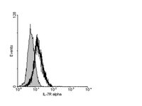
Flow Cytometry Analysis: 5 µg/mL from a previous lot detected IL-7Ra in lymphocytes.
ELISA: A previous lot of this antibody was confirmed by ELISA by an independent laboratory.
Biological Information
Immunogen
KLH-conjugated linear peptide corresponding to human IL-7Ra at and around the extracellular domain.
Epitope
Extracellular Domain
Concentration
Please refer to the Certificate of Analysis for the lot-specific concentration.
Host
Rabbit
Specificity
This antibody recognizes the extracellular domain of IL-7Ra. May detect various isoforms of IL-7Ra (see molecular weight).
The protein encoded by this gene is a receptor for interleukin 7 (IL7). The function of this receptor requires the interleukin 2 receptor, gamma chain (IL2RG), which is a common gamma chain shared by the receptors of various cytokines, including interleukin 2, 4, 7, 9, and 15. This protein has been shown to play a critical role in the V(D)J recombination during lymphocyte development. This protein is also found to control the accessibility of the TCR gamma locus by STAT5 and histone acetylation. Knockout studies in mice suggested that blocking apoptosis is an essential function of this protein during differentiation and activation of T lymphocytes. The functional defects in this protein may be associated with the pathogenesis of the severe combined immunodeficiency (SCID). [provided by RefSeq]
FUNCTION: Receptor for interleukin-7. Also acts as a receptor for thymic stromal lymphopoietin (TSLP).
SUBUNIT STRUCTURE: The IL7 receptor is an heterodimer of IL7R and IL2RG. The TSLP receptor is an heterodimer of CRLF2 and IL7R.
SUBCELLULAR LOCATION: Isoform 1: Cell membrane; Single-pass type I membrane protein.
Isoform 3: Cell membrane; Single-pass type I membrane protein.
Isoform 4: Secreted.
DOMAIN: The WSXWS motif appears to be necessary for proper protein folding and thereby efficient intracellular transport and cell-surface receptor binding.
The box 1 motif is required for JAK interaction and/or activation.
INVOLVEMENT IN DISEASE: Defects in IL7R are a cause of autosomal recessive severe combined immunodeficiency T-cell-negative/B-cell-positive/NK cell-positive (T-/B+/NK+ SCID) [MIM:608971]. SCID refers to a genetically and clinically group of rare congenital disorders characterized by impairment of both humoral and cell-mediated immunity, leukopenia, and low or absent antibody levels. Patients with SCID present in infancy with recurrent, persistent infections by opportunistic organisms, including Candida albicans, Pneumocystis carinii, and cytomegalovirus, among many others. The common characteristic of all types of SCID is absence of T-cell-mediated cellular immunity due to a defect in T-cell development.
A genetic variation in transmembrane domain of IL7R is associated with susceptibility to multiple sclerosis (MS) [MIM:126200]. Overtransmission of the major 'C' allele coding for Thr-244 are detected in offspring affected with multiple sclerosis. In vitro analysis of transcripts from minigenes containing either 'C' allele (Thr-244) or 'T' allele (Ile-244) shows that the 'C' allele results in an approximately two-fold increase in the skipping of exon 6, leading to increased production of a soluble form of IL7R. Thus, the multiple sclerosis associated 'C' risk allele of IL7R would probably decrease membrane-bound expression of IL7R. As this risk allele is common in the general population, some additional triggers are probably required for the development and progression of MS.
SEQUENCE SIMILARITIES: Belongs to the type I cytokine receptor family. Type 4 subfamily.
Contains 1 fibronectin type-III domain.
Molecular Weight
~52 kDa. May detect other isoforms of IL-7Ra produced by alternative splicing: isoform 1 (~52 kDa), isoform 2 (~29 kDa), isoform 3 (~34 kDa), and isoform 4 (~30 kDa).
Physicochemical Information
Dimensions
Materials Information
Toxicological Information
Safety Information according to GHS
Safety Information
Product Usage Statements
Quality Assurance
Evaluated by Western Blot in K562 cell lysate.
Western Blot Analysis: 1 µg/mL of this antibody detected IL-7Ra on 10 µg of K562 cell lysate.
Usage Statement
Unless otherwise stated in our catalog or other company documentation accompanying the product(s), our products are intended for research use only and are not to be used for any other purpose, which includes but is not limited to, unauthorized commercial uses, in vitro diagnostic uses, ex vivo or in vivo therapeutic uses or any type of consumption or application to humans or animals.