Wenn Sie das Fenster schließen, wird Ihre Konfiguration nicht gespeichert, es sei denn, Sie haben Ihren Artikel in die Bestellung aufgenommen oder zu Ihren Favoriten hinzugefügt.
Klicken Sie auf OK, um das MILLIPLEX® MAP-Tool zu schließen oder auf Abbrechen, um zu Ihrer Auswahl zurückzukehren.
Wählen Sie konfigurierbare Panels & Premixed-Kits - ODER - Kits für die zelluläre Signaltransduktion & MAPmates™
Konfigurieren Sie Ihre MILLIPLEX® MAP-Kits und lassen sich den Preis anzeigen.
Konfigurierbare Panels & Premixed-Kits
Unser breites Angebot enthält Multiplex-Panels, für die Sie die Analyten auswählen können, die am besten für Ihre Anwendung geeignet sind. Unter einem separaten Register können Sie das Premixed-Cytokin-Format oder ein Singleplex-Kit wählen.
Kits für die zelluläre Signaltransduktion & MAPmates™
Wählen Sie gebrauchsfertige Kits zur Erforschung gesamter Signalwege oder Prozesse. Oder konfigurieren Sie Ihre eigenen Kits mit Singleplex MAPmates™.
Die folgenden MAPmates™ sollten nicht zusammen analysiert werden: -MAPmates™, die einen unterschiedlichen Assaypuffer erfordern. -Phosphospezifische und MAPmate™ Gesamtkombinationen wie Gesamt-GSK3β und Gesamt-GSK3β (Ser 9). -PanTyr und locusspezifische MAPmates™, z.B. Phospho-EGF-Rezeptor und Phospho-STAT1 (Tyr701). -Mehr als 1 Phospho-MAPmate™ für ein einziges Target (Akt, STAT3). -GAPDH und β-Tubulin können nicht mit Kits oder MAPmates™, die panTyr enthalten, analysiert werden.
.
Bestellnummer
Bestellinformationen
St./Pkg.
Liste
Dieser Artikel wurde zu Ihren Favoriten hinzugefügt.
Wählen Sie bitte Spezies, Panelart, Kit oder Probenart
Um Ihr MILLIPLEX® MAP-Kit zu konfigurieren, wählen Sie zunächst eine Spezies, eine Panelart und/oder ein Kit.
Custom Premix Selecting "Custom Premix" option means that all of the beads you have chosen will be premixed in manufacturing before the kit is sent to you.
Catalogue Number
Ordering Description
Qty/Pack
List
Dieser Artikel wurde zu Ihren Favoriten hinzugefügt.
Spezies
Panelart
Gewähltes Kit
Menge
Bestellnummer
Bestellinformationen
St./Pkg.
Listenpreis
96-Well Plate
Menge
Bestellnummer
Bestellinformationen
St./Pkg.
Listenpreis
Weitere Reagenzien hinzufügen (MAPmates erfordern die Verwendung eines Puffer- und Detektionskits)
Menge
Bestellnummer
Bestellinformationen
St./Pkg.
Listenpreis
48-602MAG
Buffer Detection Kit for Magnetic Beads
1 Kit
Platzsparende Option Kunden, die mehrere Kits kaufen, können ihre Multiplex-Assaykomponenten in Kunststoffbeuteln anstelle von Packungen erhalten, um eine kompaktere Lagerung zu ermöglichen.
Dieser Artikel wurde zu Ihren Favoriten hinzugefügt.
Das Produkt wurde in Ihre Bestellung aufgenommen
Sie können nun ein weiteres Kit konfigurieren, ein Premixed-Kit wählen, zur Kasse gehen oder das Bestell-Tool schließen.
Detect HMG CoA Reductase using this mouse monoclonal Anti-HMG-CoA Reductase, clone IgG-A9, Cat. No. MABS1233, validated for use in Immunocytochemistry, Immunoprecipitation, and Western Blotting.
More>>Detect HMG CoA Reductase using this mouse monoclonal Anti-HMG-CoA Reductase, clone IgG-A9, Cat. No. MABS1233, validated for use in Immunocytochemistry, Immunoprecipitation, and Western Blotting. Less<<
SDB (Sicherheitsdatenblätter), Analysenzertifikate und Qualitätszertifikate, Dossiers, Broschüren und andere verfügbare Dokumente.
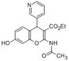
3-hydroxy-3-methylglutaryl-coenzyme A reductase (EC 1.1.1.34; UniProt P00347; also known as HMG-CoA reductase) is encoded by the HMGCR gene (Gene ID 100756363) in Cricetulus griseus (Chinese hamster). HMG-CoA reductase catalyzes the conversion of HMG-CoA to mevalonic acid, the rate-limiting enzyme of the mevalonate-isoprenoid biosynthesis (MIB) pathway responsible for the production of cholesterol and other isoprenoids, as well as farnesyl pyrophosphate (FPP) required for protein prenylation. HMG-CoA reductase is suppressed by cholesterol derived from the internalization and degradation of low density lipoprotein (LDL) via the LDL receptor as well as oxidized cholesterol species. Competitive inhibitors against HMG-CoA reductase induce LDL receptors expression in the liver, which in turn increases the catabolism of plasma LDL and lowers the plasma concentration of cholesterol. HMG-CoA reductase is a multitransmembrane ER protein with an N-terminal sterol-sensing domain (SSD; a.a. 61-218) and a C-terminal catalytic domain (a.a. 450-888) whose activity is inhibited upon SSD binding by cholesterol. Numerous studies have implicated the involvement of HMG-CoA reductase and mevalonate pathway in carcinogenesis.
References
Product Information
Format
Purified
Presentation
Purified mouse IgG1 in buffer containing 0.1 M Tris-Glycine (pH 7.4), 150 mM NaCl with 0.05% sodium azide.
Detect HMG CoA Reductase using this mouse monoclonal Anti-HMG-CoA Reductase, clone IgG-A9, Cat. No. MABS1233, validated for use in Immunocytochemistry, Immunoprecipitation, and Western Blotting.
Key Applications
Immunocytochemistry
Western Blotting
Immunoprecipitation
Application Notes
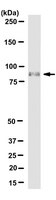
Western Blotting Analysis: A 1:100 dilution from a representative lot detected in 15 µg of membrane extract from CHO7 cells treated overnight with 0.01 mM compactin (Courtesy of Linda Donnelly, Department of Molecular Genetics, UT Southwestern Medical Center, Dallas, TX, USA).
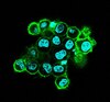
Immunocytochemistry Analysis: A representative lot detected HMG CoA reductase-positive ER membrane structures by indirect immunofluorescence staining of paraformaldehyde-fixed, 0.1% Triton X-100-permeabilized CHO-K1 cells cultured with compactin, mevalonate, and MG-132, in the presence or absence of 25-hydroxycholesterol (25-HC) (Hartman, I.Z., et al. (2010). J. Biol. Chem. 285(25):19288-19298).
Immunoprecipitation Analysis: A representative lot immunodepleted HMC-CoA reductase activity from compactin-adapted CHO cell (UT-1) extract (Liscum, L., et al. (1983). J. Biol. Chem. 258(13) 8450-8455).
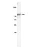
Western Blotting Analysis: A representative lot detected 25-hydroxycholesterol (25-HC) treatment-induced HMC-CoA reductase dislocation from ER membrane to the cytosol and lipid droplet in CHO-K1 cells cultured with lipoprotein-deficient serum in the presence of compactin, MG-132, with or without mevalonate. Cellular VCP knockdown prevented sterol-induced HMC-CoA reductase cytosolic, but not lipid droplet, translocation (Hartman, I.Z., et al. (2010). J. Biol. Chem. 285(25):19288-19298).
Western Blotting Analysis: A representative lot detected 25-hydroxycholesterol (25-HC) treatment-induced ubiquitination of HMC-CoA reductase in SV-589 human fibroblasts cultured with compactin and mevalonate in the presence of proteasome inhibitor MG-132. Cellular knockdown of gp78A, but not gp78B or Hrd1, reduced 25-HC-induced HMC-CoA reductase ubiquitination (Song, B.L., et al. (2005). Mol. Cell. 19(6):829-840).
Western Blotting Analysis: A representative lot detected 25-hydroxycholesterol (25-HC) treatment-induced degradation of HMC-CoA reductase in SV-589 human fibroblasts cultured with compactin and mevalonate. Cellular knockdown of gp78A, VCP, or Insig-1/-2 suppressed sterol-induced HMC-CoA reductase degradation (Song, B.L., et al. (2005). Mol. Cell. 19(6):829-840).
Western Blotting Analysis: A representative lot detected HMC-CoA reductase in both membrane and nuclear fractions from HEK-293S cells cultured with compactin and mevalonate. Additional treatment with 25-hydroxycholesterol (25-HC) resulted in HMC-CoA reductase degradation (Sever, N., et al. (2003). Mol. Cell. 11(1):25-33).
Western Blotting Analysis: A representative lot detected an upregulated HMC-CoA reductase expression in compactin-adapted CHO cells (UT-1), as well as a loss of HMC-CoA reductase expression in UT-1 cells cultured in the presence of LDL (Liscum, L., et al. (1983). J. Biol. Chem. 258(13) 8450-8455).
Western Blotting Analysis: A representative lot detected a greater HMC-CoA reductase upregulation in liver microsome preparation from rats on diet supplemented with cholestyramine and mevinolin than with cholestyramine alone, while supplementation with cholesterol in addition to cholestyramine and mevinolin downregulated liver HMC-CoA reductase (Liscum, L., et al. (1983). J. Biol. Chem. 258(13) 8450-8455).
Biological Information
Immunogen
Membrane preapration from compactin-adapted CHO cells (Liscum, L., et al. (1983). J. Biol. Chem. 258(13) 8450-8455).
Clone
IgG-A9
Concentration
Please refer to lot specific datasheet.
Host
Mouse
Specificity
Clone IgG-A9 targets an epitope within HMC-CoA reductase catalytic domain (a.a. 450 887).
~92 kDa observed. 97.08 kDa (hamster), 97.48/92.02/99.75 kDa (human isoform 1/2/3 (HMGCR-1b)), 96.69 kDa (rat) calculated.. Uncharacterized bands may be observed in some lysate(s).
Physicochemical Information
Dimensions
Materials Information
Toxicological Information
Safety Information according to GHS
Safety Information
Product Usage Statements
Quality Assurance
Identity Confirmation by Isotyping Test.
Isotyping Analysis: The identity of this monoclonal antibody is confirmed by isotyping test to be mouse IgG1 .
Usage Statement
Unless otherwise stated in our catalog or other company documentation accompanying the product(s), our products are intended for research use only and are not to be used for any other purpose, which includes but is not limited to, unauthorized commercial uses, in vitro diagnostic uses, ex vivo or in vivo therapeutic uses or any type of consumption or application to humans or animals.