Wenn Sie das Fenster schließen, wird Ihre Konfiguration nicht gespeichert, es sei denn, Sie haben Ihren Artikel in die Bestellung aufgenommen oder zu Ihren Favoriten hinzugefügt.
Klicken Sie auf OK, um das MILLIPLEX® MAP-Tool zu schließen oder auf Abbrechen, um zu Ihrer Auswahl zurückzukehren.
Wählen Sie konfigurierbare Panels & Premixed-Kits - ODER - Kits für die zelluläre Signaltransduktion & MAPmates™
Konfigurieren Sie Ihre MILLIPLEX® MAP-Kits und lassen sich den Preis anzeigen.
Konfigurierbare Panels & Premixed-Kits
Unser breites Angebot enthält Multiplex-Panels, für die Sie die Analyten auswählen können, die am besten für Ihre Anwendung geeignet sind. Unter einem separaten Register können Sie das Premixed-Cytokin-Format oder ein Singleplex-Kit wählen.
Kits für die zelluläre Signaltransduktion & MAPmates™
Wählen Sie gebrauchsfertige Kits zur Erforschung gesamter Signalwege oder Prozesse. Oder konfigurieren Sie Ihre eigenen Kits mit Singleplex MAPmates™.
Die folgenden MAPmates™ sollten nicht zusammen analysiert werden: -MAPmates™, die einen unterschiedlichen Assaypuffer erfordern. -Phosphospezifische und MAPmate™ Gesamtkombinationen wie Gesamt-GSK3β und Gesamt-GSK3β (Ser 9). -PanTyr und locusspezifische MAPmates™, z.B. Phospho-EGF-Rezeptor und Phospho-STAT1 (Tyr701). -Mehr als 1 Phospho-MAPmate™ für ein einziges Target (Akt, STAT3). -GAPDH und β-Tubulin können nicht mit Kits oder MAPmates™, die panTyr enthalten, analysiert werden.
.
Bestellnummer
Bestellinformationen
St./Pkg.
Liste
Dieser Artikel wurde zu Ihren Favoriten hinzugefügt.
Wählen Sie bitte Spezies, Panelart, Kit oder Probenart
Um Ihr MILLIPLEX® MAP-Kit zu konfigurieren, wählen Sie zunächst eine Spezies, eine Panelart und/oder ein Kit.
Custom Premix Selecting "Custom Premix" option means that all of the beads you have chosen will be premixed in manufacturing before the kit is sent to you.
Catalogue Number
Ordering Description
Qty/Pack
List
Dieser Artikel wurde zu Ihren Favoriten hinzugefügt.
Spezies
Panelart
Gewähltes Kit
Menge
Bestellnummer
Bestellinformationen
St./Pkg.
Listenpreis
96-Well Plate
Menge
Bestellnummer
Bestellinformationen
St./Pkg.
Listenpreis
Weitere Reagenzien hinzufügen (MAPmates erfordern die Verwendung eines Puffer- und Detektionskits)
Menge
Bestellnummer
Bestellinformationen
St./Pkg.
Listenpreis
48-602MAG
Buffer Detection Kit for Magnetic Beads
1 Kit
Platzsparende Option Kunden, die mehrere Kits kaufen, können ihre Multiplex-Assaykomponenten in Kunststoffbeuteln anstelle von Packungen erhalten, um eine kompaktere Lagerung zu ermöglichen.
Dieser Artikel wurde zu Ihren Favoriten hinzugefügt.
Das Produkt wurde in Ihre Bestellung aufgenommen
Sie können nun ein weiteres Kit konfigurieren, ein Premixed-Kit wählen, zur Kasse gehen oder das Bestell-Tool schließen.
Anti-GCAP1, clone 6B12, Cat. No. MABN2396, is a highly specific mouse monoclonal antibody that targets Guanylyl cyclase-activating protein 1 and has been tested in Immunofluorescence, Immunohistochemistry (Paraffin), Immunoprecipitation, and Western Blotting.
More>>Anti-GCAP1, clone 6B12, Cat. No. MABN2396, is a highly specific mouse monoclonal antibody that targets Guanylyl cyclase-activating protein 1 and has been tested in Immunofluorescence, Immunohistochemistry (Paraffin), Immunoprecipitation, and Western Blotting. Less<<
Anti-GCAP1 Antibody, clone 6B12: SDB (Sicherheitsdatenblätter), Analysenzertifikate und Qualitätszertifikate, Dossiers, Broschüren und andere verfügbare Dokumente.
Guanylyl cyclase-activating protein 1 (UniProt: P43081; also known as GCAP 1, Guanylate cyclase activator 1A) is encoded by the Guca1a (also known as Gcap, Gcap1, Guca1) gene (Gene ID: 14913) in murine species. GCAP1 is a membrane bound protein that stimulates retinal guanylyl cyclase when free calcium ions concentration is low and inhibits guanylyl cyclase when free calcium ions concentration is elevated. This Ca2+-sensitive regulation of retinal guanylyl cyclase appears to be a key event in recovery of the dark state of rod photoreceptors following light exposure. GCAP1 is present in rod and cone photoreceptor outer segments where phototransduction occurs. It binds three calcium ions (via EF-hands 2, 3 and 4) when calcium levels are high. Binds Mg2+ when calcium levels are low. Three calcium-binding sites are identified (aa 64-75; 100-111; and 144-155). GCAP1 restores the calcium sensitivity of guanylyl cyclase in a reconstituted system, and it decreases the sensitivity, time-to-peak, and recovery time of the light response following its introduction into intact rod outer segments. (Ref.: Gorczyca WA (1995). J. Biol Chem. 270(37); 22029-36; Schrem A et al. (1999). J Biol Chem. 274(10); 6244-49).
References
Product Information
Format
Purified
Presentation
Purified mouse monoclonal antibody IgG2a in buffer containing 0.1 M Tris-Glycine (pH 7.4), 150 mM NaCl with 0.05% sodium azide.
Anti-GCAP1, clone 6B12, Cat. No. MABN2396, is a highly specific mouse monoclonal antibody that targets Guanylyl cyclase-activating protein 1 and has been tested in Immunofluorescence, Immunohistochemistry (Paraffin), Immunoprecipitation, and Western Blotting.
Key Applications
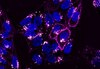
Immunofluorescence
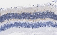
Immunohistochemistry (Paraffin)
Immunoprecipitation
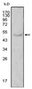
Western Blotting
Application Notes
Immunofluorescence Analysis: A representative lot detected GCAP1 in Immunofluorescence applications (Zulliger, R., et. al. (2015). J Biol Chem. 290(6):3488-99).
Immunofluorescence Analysis: A 1:10 dilution from a representative lot detected GCAP1 in mouse retina.
Western Blotting Analysis: A representative lot detected GCAP1 in Western Blotting applications (Zulliger, R., et. al. (2015). J Biol Chem. 290(6):3488-99).
Immunoprecipitation Analysis: A representative lot detected GCAP1 in Immunoprecipitation applications (Zulliger, R., et. al. (2015). J Biol Chem. 290(6):3488-99).
Immunohistochemistry Analysis: A 1:50 dilution from a representative lot detected GCAP1 in human retina tissue.
Biological Information
Immunogen
GST-fusion protein containing 45 amino acids from the C-terminal region of murine GCAP1. It does not display any homology with GCAP2.
Epitope
C-terminus
Clone
6B12
Concentration
Please refer to lot specific datasheet.
Host
Mouse
Specificity
Clone 6B12 detects Guanylyl cyclase-activating protein 1 in Bovine, Human, Mouse, Porcine, and Rat. It targets an epitope within 45 amino acids from the C-terminal region.
23 kDa observed; 22.99 kDa calculated. Uncharacterized bands may be observed in some lysate(s).
Physicochemical Information
Dimensions
Materials Information
Toxicological Information
Safety Information according to GHS
Safety Information
Product Usage Statements
Quality Assurance
Evaluated by Western Blotting in C57BL/6 mouse retinal tissue lysate.
Western Blotting Analysis: 1 µg/mL of this antibody detected GCAP1 in C57BL/6 mouse retinal tissue lysate.
Usage Statement
Unless otherwise stated in our catalog or other company documentation accompanying the product(s), our products are intended for research use only and are not to be used for any other purpose, which includes but is not limited to, unauthorized commercial uses, in vitro diagnostic uses, ex vivo or in vivo therapeutic uses or any type of consumption or application to humans or animals.