Wenn Sie das Fenster schließen, wird Ihre Konfiguration nicht gespeichert, es sei denn, Sie haben Ihren Artikel in die Bestellung aufgenommen oder zu Ihren Favoriten hinzugefügt.
Klicken Sie auf OK, um das MILLIPLEX® MAP-Tool zu schließen oder auf Abbrechen, um zu Ihrer Auswahl zurückzukehren.
Wählen Sie konfigurierbare Panels & Premixed-Kits - ODER - Kits für die zelluläre Signaltransduktion & MAPmates™
Konfigurieren Sie Ihre MILLIPLEX® MAP-Kits und lassen sich den Preis anzeigen.
Konfigurierbare Panels & Premixed-Kits
Unser breites Angebot enthält Multiplex-Panels, für die Sie die Analyten auswählen können, die am besten für Ihre Anwendung geeignet sind. Unter einem separaten Register können Sie das Premixed-Cytokin-Format oder ein Singleplex-Kit wählen.
Kits für die zelluläre Signaltransduktion & MAPmates™
Wählen Sie gebrauchsfertige Kits zur Erforschung gesamter Signalwege oder Prozesse. Oder konfigurieren Sie Ihre eigenen Kits mit Singleplex MAPmates™.
Die folgenden MAPmates™ sollten nicht zusammen analysiert werden: -MAPmates™, die einen unterschiedlichen Assaypuffer erfordern. -Phosphospezifische und MAPmate™ Gesamtkombinationen wie Gesamt-GSK3β und Gesamt-GSK3β (Ser 9). -PanTyr und locusspezifische MAPmates™, z.B. Phospho-EGF-Rezeptor und Phospho-STAT1 (Tyr701). -Mehr als 1 Phospho-MAPmate™ für ein einziges Target (Akt, STAT3). -GAPDH und β-Tubulin können nicht mit Kits oder MAPmates™, die panTyr enthalten, analysiert werden.
.
Bestellnummer
Bestellinformationen
St./Pkg.
Liste
Dieser Artikel wurde zu Ihren Favoriten hinzugefügt.
Wählen Sie bitte Spezies, Panelart, Kit oder Probenart
Um Ihr MILLIPLEX® MAP-Kit zu konfigurieren, wählen Sie zunächst eine Spezies, eine Panelart und/oder ein Kit.
Custom Premix Selecting "Custom Premix" option means that all of the beads you have chosen will be premixed in manufacturing before the kit is sent to you.
Catalogue Number
Ordering Description
Qty/Pack
List
Dieser Artikel wurde zu Ihren Favoriten hinzugefügt.
Spezies
Panelart
Gewähltes Kit
Menge
Bestellnummer
Bestellinformationen
St./Pkg.
Listenpreis
96-Well Plate
Menge
Bestellnummer
Bestellinformationen
St./Pkg.
Listenpreis
Weitere Reagenzien hinzufügen (MAPmates erfordern die Verwendung eines Puffer- und Detektionskits)
Menge
Bestellnummer
Bestellinformationen
St./Pkg.
Listenpreis
48-602MAG
Buffer Detection Kit for Magnetic Beads
1 Kit
Platzsparende Option Kunden, die mehrere Kits kaufen, können ihre Multiplex-Assaykomponenten in Kunststoffbeuteln anstelle von Packungen erhalten, um eine kompaktere Lagerung zu ermöglichen.
Dieser Artikel wurde zu Ihren Favoriten hinzugefügt.
Das Produkt wurde in Ihre Bestellung aufgenommen
Sie können nun ein weiteres Kit konfigurieren, ein Premixed-Kit wählen, zur Kasse gehen oder das Bestell-Tool schließen.
ABC474
Sigma-AldrichAnti-Fhl1 Antibody
Detect Four & a half LIM domains protein 1 using this rabbit polyclonal antibody, Anti-Fhl1 Antibody validated for use in western blotting, ICC & IHC.
More>>Detect Four & a half LIM domains protein 1 using this rabbit polyclonal antibody, Anti-Fhl1 Antibody validated for use in western blotting, ICC & IHC. Less<<
Anti-Fhl1 Antibody: SDB (Sicherheitsdatenblätter), Analysenzertifikate und Qualitätszertifikate, Dossiers, Broschüren und andere verfügbare Dokumente.
Four and a half LIM domains protein 1 (FHL1) is also called Skeletal muscle LIM-protein 1 (SLIM-1). FHL1 has 3 isoforms; Isoform 1 is highly expressed in skeletal muscle; Isoform 2 is expressed in brain and skeletal muscle; and Isoform 3 is expressed in testis, heart and skeletal muscle. FHL1 is possibly involved in muscle development or hypertrophy and associated with X-linked dominant scapuloperoneal myopathy (SPM), X-linked myopathy with postural muscle atrophy (XMPMA), X-linked severe early-onset reducing body myopathy (RBM) and X-linked childhood-onset reducing body myopathy (CO-RBM).
References
Product Information
Format
Serum
Presentation
Rabbit polyclonal in buffer containing serum with 0.05% sodium azide.
Detect Four & a half LIM domains protein 1 using this rabbit polyclonal antibody, Anti-Fhl1 Antibody validated for use in western blotting, ICC & IHC.
Key Applications
Western Blotting
Immunocytochemistry
Immunohistochemistry
Application Notes
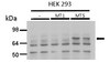
Western Blotting Analysis: A 1:1,000 dilution from a representative lot detected Fhl1 in a panel of select tissue lysates.
Western Blotting Analysis: A 1:1,000 dilution from a representative lot detected Fhl1 in an additional panel of select tissue lysates.
Western Blotting Analysis: A representative lot from an independent laboratory detected Fhl1 in nonanchored and anchored CasKO and Cx43KO cell lysates. A representative lot from the same independent laboratory detected Fhl1 in non-SRC transformed Cx43KO cell lysate and not in SRC transformed Cx43KO cell lysate (Shen, Y., et al. (2006). Cancer Res. 66(3):1543-1552.).
Western Blotting Analysis: A representative lot from an independent laboratory detected Fhl1 in non-SRC transformed Cx43KO cell lysate and not in SRC transformed Cx43KO cell lysate (Li, X., et al. (2008). Cancer Sci. 99(7):1326-1333.).
Immunohistochemistry Analysis: A representative lot from an independent laboratory detected Fhl1 in normal brain, kidney, liver, lung, prostate, and skin tissues and demonstrated a loss of signal in breast carcinoma, renal carcinoma, hepatocarcinoma, pulmonary carcinoma, prostatic carcinoma, and melanoma tissues (Shen, Y., et al. (2006). Cancer Res. 66(3):1543-1552.).
Immunohistochemistry Analysis: A representative lot from an independent laboratory detected Fhl1 in normal epithelium of breast, kidney, and prostate tissues, and demonstrated a loss of signal in mammary duct carcinoma, renal cell carcinoma, and prostate adenocarcinoma tissues (Li, X., et al. (2008). Cancer Sci. 99(7):1326-1333.).
Immunocytochemistry Analysis: A representative lot from an independent laboratory detected Fhl1 in non SRC transformed Cx43KO and CasKO cells and demonstrated a loss of signal in SRC transformed Cx43KO and CasKO cells (Shen, Y., et al. (2006). Cancer Res. 66(3):1543-1552.).
Biological Information
Immunogen
Linear peptide corresponding to amino acid sequence CRDPLQGKKYVQKDGRH of human Fhl1.
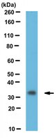
~32 kDa observed. The calculated molecular weight of isoform 1 for this protein is 32 kDa; however, a dimer may be observed at ~64 kDa in some cell lysates. Uncharacterized band(s) may be observed in some cell lysates.
Physicochemical Information
Dimensions
Materials Information
Toxicological Information
Safety Information according to GHS
Safety Information
Product Usage Statements
Quality Assurance
Evaluated by Western Blotting in human brain tissue lysate.
Western Blotting Analysis: A 1:1,000 dilution of this antibody detected Fhl1 in 10 µg of human brain tissue lysate.
Usage Statement
Unless otherwise stated in our catalog or other company documentation accompanying the product(s), our products are intended for research use only and are not to be used for any other purpose, which includes but is not limited to, unauthorized commercial uses, in vitro diagnostic uses, ex vivo or in vivo therapeutic uses or any type of consumption or application to humans or animals.
Storage and Shipping Information
Storage Conditions
Stable for 1 year at -20°C from date of receipt. Handling Recommendations: Upon receipt and prior to removing the cap, centrifuge the vial and gently mix the solution. Aliquot into microcentrifuge tubes and store at -20°C. Avoid repeated freeze/thaw cycles, which may damage IgG and affect product performance.
SRC uses Cas to suppress Fhl1 in order to promote nonanchored growth and migration of tumor cells. Shen, Yongquan, et al. Cancer Res., 66: 1543-52 (2006)
2005
Anchorage independence and motility are hallmarks of tumor cell growth. Tumor cell growth and morphology can be normalized by contact with nontransformed cells. The Src tyrosine kinase phosphorylates specific sites on the focal adhesion adaptor protein Crk-associated substrate (Cas) to promote nonanchored cell growth and migration. We studied the effects of Src and Cas on the expression of >14,000 genes to identify molecular events that underlie these activities. Gene expression in tumor cells that were normalized by neighboring nontransformed cells was used as an additional filter to identify genes that control metastatic cell growth. This process enabled the identification of genes that play roles in anchorage-independent cell growth and migration. One candidate, four and a half LIM domains 1 (Fhl1), acts as a transcriptional regulator that can associate with cell junctions as well as with the nucleus. We show here that Src phosphorylates Cas to block Fhl1 expression. In addition, suppression of Fhl1 is required for Src to promote tumor cell growth. These data show that Fhl1 is a tumor suppressor gene that acts downstream of Src and Cas to specifically block anchorage-independent cell growth and migration. Moreover, Fhl1 was suppressed in tumors from several human tissues. Thus, identification of how Fhl1 controls fundamental aspects of tumor cell growth and metastasis may lead to the development of novel markers that can be used to diagnose human clinical specimens as well as open innovative avenues of investigations aimed at developing reagents that target cancer cells while minimizing damage to normal cells.