Wenn Sie das Fenster schließen, wird Ihre Konfiguration nicht gespeichert, es sei denn, Sie haben Ihren Artikel in die Bestellung aufgenommen oder zu Ihren Favoriten hinzugefügt.
Klicken Sie auf OK, um das MILLIPLEX® MAP-Tool zu schließen oder auf Abbrechen, um zu Ihrer Auswahl zurückzukehren.
Wählen Sie konfigurierbare Panels & Premixed-Kits - ODER - Kits für die zelluläre Signaltransduktion & MAPmates™
Konfigurieren Sie Ihre MILLIPLEX® MAP-Kits und lassen sich den Preis anzeigen.
Konfigurierbare Panels & Premixed-Kits
Unser breites Angebot enthält Multiplex-Panels, für die Sie die Analyten auswählen können, die am besten für Ihre Anwendung geeignet sind. Unter einem separaten Register können Sie das Premixed-Cytokin-Format oder ein Singleplex-Kit wählen.
Kits für die zelluläre Signaltransduktion & MAPmates™
Wählen Sie gebrauchsfertige Kits zur Erforschung gesamter Signalwege oder Prozesse. Oder konfigurieren Sie Ihre eigenen Kits mit Singleplex MAPmates™.
Die folgenden MAPmates™ sollten nicht zusammen analysiert werden: -MAPmates™, die einen unterschiedlichen Assaypuffer erfordern. -Phosphospezifische und MAPmate™ Gesamtkombinationen wie Gesamt-GSK3β und Gesamt-GSK3β (Ser 9). -PanTyr und locusspezifische MAPmates™, z.B. Phospho-EGF-Rezeptor und Phospho-STAT1 (Tyr701). -Mehr als 1 Phospho-MAPmate™ für ein einziges Target (Akt, STAT3). -GAPDH und β-Tubulin können nicht mit Kits oder MAPmates™, die panTyr enthalten, analysiert werden.
.
Bestellnummer
Bestellinformationen
St./Pkg.
Liste
Dieser Artikel wurde zu Ihren Favoriten hinzugefügt.
Wählen Sie bitte Spezies, Panelart, Kit oder Probenart
Um Ihr MILLIPLEX® MAP-Kit zu konfigurieren, wählen Sie zunächst eine Spezies, eine Panelart und/oder ein Kit.
Custom Premix Selecting "Custom Premix" option means that all of the beads you have chosen will be premixed in manufacturing before the kit is sent to you.
Catalogue Number
Ordering Description
Qty/Pack
List
Dieser Artikel wurde zu Ihren Favoriten hinzugefügt.
Spezies
Panelart
Gewähltes Kit
Menge
Bestellnummer
Bestellinformationen
St./Pkg.
Listenpreis
96-Well Plate
Menge
Bestellnummer
Bestellinformationen
St./Pkg.
Listenpreis
Weitere Reagenzien hinzufügen (MAPmates erfordern die Verwendung eines Puffer- und Detektionskits)
Menge
Bestellnummer
Bestellinformationen
St./Pkg.
Listenpreis
48-602MAG
Buffer Detection Kit for Magnetic Beads
1 Kit
Platzsparende Option Kunden, die mehrere Kits kaufen, können ihre Multiplex-Assaykomponenten in Kunststoffbeuteln anstelle von Packungen erhalten, um eine kompaktere Lagerung zu ermöglichen.
Dieser Artikel wurde zu Ihren Favoriten hinzugefügt.
Das Produkt wurde in Ihre Bestellung aufgenommen
Sie können nun ein weiteres Kit konfigurieren, ein Premixed-Kit wählen, zur Kasse gehen oder das Bestell-Tool schließen.
Anti-ERR alpha, clone 2ERR10, Cat. No. MABE1891, is a mouse monoclonal antibody that detects Steroid hormone receptor ERR alpha and has been tested for use in Chromatin Immunoprecipitation, ELISA, Immunocytochemistry, Immunoprecipitation, and Western Blotting.
More>>Anti-ERR alpha, clone 2ERR10, Cat. No. MABE1891, is a mouse monoclonal antibody that detects Steroid hormone receptor ERR alpha and has been tested for use in Chromatin Immunoprecipitation, ELISA, Immunocytochemistry, Immunoprecipitation, and Western Blotting. Less<<
Empfohlene Produkte
Übersicht
Replacement Information
Description
Catalogue Number
MABE1891-100UG
Description
Anti-ERR alpha Antibody, clone 2ERR10
Alternate Names
Steroid hormone receptor ERR1
Estrogen receptor-like 1
Estrogen-related receptor alpha
Nuclear receptor subfamily 3 group B member 1
Background Information
Steroid hormone receptor ERR1 (UniProt: P11474; also known as Estrogen receptor-like 1, Estrogen-related receptor alpha, ERR-alpha, Nuclear receptor subfamily 3 group B member 1) is encoded by the ESRRA (also known as ERR1, ESRL1, NR3B1) gene (Gene ID: 2101) in human. ERR1 is a orphan nuclear receptor with no known natural ligands. Its transcriptional activities are thought to be modulated by post-translational modifications and interactions with cofactors. It is localized predominantly in cell nuclei but can co-localize to the cytoplasm in presence of MAPK15. It is most highly expressed in skeletal muscle, kidney, heart, brain, and intestine. ERR1 contains four major canonical nuclear receptor domains. The N-terminal A/B domain (aa 1-78) is involved in ligand-independent functions of nuclear receptors. The C region (aa 79-144) contains the DNA-binding domain (DBD)and adjacent to the DBD is the D domain (aa 145-198), which is also known as the hinge region. The C-terminal E/F domain (aa 199-423) typically contains a ligand-binding region for most nuclear receptors. ERR1 contains two NR C-type zinc finger domains (aa 79-99 and 115-134). Its activity is induced by PGC1 alpha in several specific cell types. ERR1 regulates genes involved in mitochondrial biogenesis, gluconeogenesis, oxidative phosphorylation, and fatty acid metabolism, and brown adipose tissue thermogenesis. Phosphorylation of ERR1 at serine 19 is shown to enhance its sumolyation on lysine 14, which can increase repression of its transcriptional activity. Over-expression of ERR1 has been linked to breast, colon, and ovarian cancers. (Ref.: Esch, AM., et al. (2012). Protein Expression and Purification 84(1); 47-58).
References
Product Information
Format
Purified
Presentation
Purified mouse monoclonal antibody IgG1 in buffer containing 0.1 M Tris-Glycine (pH 7.4), 150 mM NaCl with 0.05% sodium azide.
Applications
Application
Anti-ERR alpha, clone 2ERR10, Cat. No. MABE1891, is a mouse monoclonal antibody that detects Steroid hormone receptor ERR alpha and has been tested for use in Chromatin Immunoprecipitation, ELISA, Immunocytochemistry, Immunoprecipitation, and Western Blotting.
Key Applications
Chromatin Immunoprecipitation (ChIP)
ELISA
Immunocytochemistry
Immunoprecipitation
Western Blotting
Application Notes
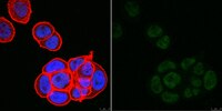
Immunocytochemistry Analysis: A 1:20 dilution from a representative lot detected ERR alpha in MCF-7 cells.
Immunoprecipitation Analysis: A representative lot immunoprecipitated ERR alpha in Immunoprecipitation applications (Esch, A.M., et. al. (2012). Protein Expr. Purif. 84(1):47-58).
Immunocytochemistry Analysis: A representative lot detected ERR alpha in Immunocytochemistry applications (Esch, A.M., et. al. (2012). Protein Expr. Purif. 84(1):47-58).
Western Blotting Analysis: A representative lot detected ERR alpha in Western Blotting applications (Esch, A.M., et. al. (2012). Protein Expr. Purif. 84(1):47-58).
Chromatin Immunoprecipitation Analysis (ChIP): A representative lot detected ERR alpha in Chromatin Immunoprecipitation applications (Esch, A.M., et. al. (2012). Protein Expr. Purrif. 84(1):47-58).
Enzyme Immunoassay Analysis: A representative lot detected ERR alpha in ELISA applications (Esch, A.M., et. al. (2012). Protein Expr. Purif. 84(1):47-58).
Biological Information
Immunogen
His-tagged full length human recombinant Steroid hormone receptor ERR alpha (ERR1).
Epitope
N-terminus
Clone
2ERR10
Concentration
Please refer to lot specific datasheet.
Host
Mouse
Specificity
Clone 2ERR10 is a mouse monoclonal antibody that detects human Steroid hormone receptor ERR alpha (ERR1).
~53 kDa observed; 45.51 kDa calculated. Uncharacterized bands may be observed in some lysate(s).
Physicochemical Information
Dimensions
Materials Information
Toxicological Information
Safety Information according to GHS
Safety Information
Product Usage Statements
Quality Assurance
Evaluated by Western Blotting in lysate from HEK293T cells transfected with ERR alpha.
Western Blotting Analysis: 1 µg/mL of this antibody detected ERR alpha in lysate from HEK293T transfected with ERR alpha.
Usage Statement
Unless otherwise stated in our catalog or other company documentation accompanying the product(s), our products are intended for research use only and are not to be used for any other purpose, which includes but is not limited to, unauthorized commercial uses, in vitro diagnostic uses, ex vivo or in vivo therapeutic uses or any type of consumption or application to humans or animals.
Production and characterization of monoclonal antibodies to estrogen-related receptor alpha (ERRα) and use in immunoaffinity chromatography. Esch, AM; Thompson, NE; Lamberski, JA; Mertz, JE; Burgess, RR Protein Expr Purif
84
47-58
2011
Estrogen-related receptor alpha (ERRα) is an orphan nuclear receptor whose elevated expression is thought to contribute to breast, colon, and ovarian cancers. In order to investigate the role of ERRα in human disease, there is a need for immunological reagents suitable for detection and purification of ERRα. We expressed recombinant human ERRα in Escherichia coli, purified the protein, and used it to generate monoclonal antibodies (mAbs) to ERRα. Nine high-affinity mAbs were chosen for their abilities to detect overexpressed ERRα in enzyme-linked immunosorbent assays (ELISAs) and Western blots, after which isotyping and preliminary epitope mapping was performed. The mAbs were all IgG subtypes and reacted with several different regions of full-length ERRα. A majority of the mAbs were found to be useful for immunoprecipitation of ERRα, and several could detect DNA-bound ERRα in electrophoretic mobility supershift assays (EMSAs) and chromatin immunoprecipitation (ChIP). The suitability of mAbs to detect ERRα in immunofluorescence assays was assessed. One mAb in particular, 2ERR10, could specifically detect endogenous ERRα in mammary carcinoma cells. Finally, we performed assays to screen for mAbs that gently release ERRα in the presence of a low-molecular-weight polyhydroxylated compound (polyol) and nonchaotropic salt. Using gentle immunoaffinity chromatography, we were able to isolate ERRα from mammalian cells by eluting with a polyol-salt solution. Our characterization studies show that these monoclonal antibodies perform well in a variety of biochemical assays. We anticipate that these novel reagents will prove useful for the detection and purification of ERRα in research and clinical applications.