Wenn Sie das Fenster schließen, wird Ihre Konfiguration nicht gespeichert, es sei denn, Sie haben Ihren Artikel in die Bestellung aufgenommen oder zu Ihren Favoriten hinzugefügt.
Klicken Sie auf OK, um das MILLIPLEX® MAP-Tool zu schließen oder auf Abbrechen, um zu Ihrer Auswahl zurückzukehren.
Wählen Sie konfigurierbare Panels & Premixed-Kits - ODER - Kits für die zelluläre Signaltransduktion & MAPmates™
Konfigurieren Sie Ihre MILLIPLEX® MAP-Kits und lassen sich den Preis anzeigen.
Konfigurierbare Panels & Premixed-Kits
Unser breites Angebot enthält Multiplex-Panels, für die Sie die Analyten auswählen können, die am besten für Ihre Anwendung geeignet sind. Unter einem separaten Register können Sie das Premixed-Cytokin-Format oder ein Singleplex-Kit wählen.
Kits für die zelluläre Signaltransduktion & MAPmates™
Wählen Sie gebrauchsfertige Kits zur Erforschung gesamter Signalwege oder Prozesse. Oder konfigurieren Sie Ihre eigenen Kits mit Singleplex MAPmates™.
Die folgenden MAPmates™ sollten nicht zusammen analysiert werden: -MAPmates™, die einen unterschiedlichen Assaypuffer erfordern. -Phosphospezifische und MAPmate™ Gesamtkombinationen wie Gesamt-GSK3β und Gesamt-GSK3β (Ser 9). -PanTyr und locusspezifische MAPmates™, z.B. Phospho-EGF-Rezeptor und Phospho-STAT1 (Tyr701). -Mehr als 1 Phospho-MAPmate™ für ein einziges Target (Akt, STAT3). -GAPDH und β-Tubulin können nicht mit Kits oder MAPmates™, die panTyr enthalten, analysiert werden.
.
Bestellnummer
Bestellinformationen
St./Pkg.
Liste
Dieser Artikel wurde zu Ihren Favoriten hinzugefügt.
Wählen Sie bitte Spezies, Panelart, Kit oder Probenart
Um Ihr MILLIPLEX® MAP-Kit zu konfigurieren, wählen Sie zunächst eine Spezies, eine Panelart und/oder ein Kit.
Custom Premix Selecting "Custom Premix" option means that all of the beads you have chosen will be premixed in manufacturing before the kit is sent to you.
Catalogue Number
Ordering Description
Qty/Pack
List
Dieser Artikel wurde zu Ihren Favoriten hinzugefügt.
Spezies
Panelart
Gewähltes Kit
Menge
Bestellnummer
Bestellinformationen
St./Pkg.
Listenpreis
96-Well Plate
Menge
Bestellnummer
Bestellinformationen
St./Pkg.
Listenpreis
Weitere Reagenzien hinzufügen (MAPmates erfordern die Verwendung eines Puffer- und Detektionskits)
Menge
Bestellnummer
Bestellinformationen
St./Pkg.
Listenpreis
48-602MAG
Buffer Detection Kit for Magnetic Beads
1 Kit
Platzsparende Option Kunden, die mehrere Kits kaufen, können ihre Multiplex-Assaykomponenten in Kunststoffbeuteln anstelle von Packungen erhalten, um eine kompaktere Lagerung zu ermöglichen.
Dieser Artikel wurde zu Ihren Favoriten hinzugefügt.
Das Produkt wurde in Ihre Bestellung aufgenommen
Sie können nun ein weiteres Kit konfigurieren, ein Premixed-Kit wählen, zur Kasse gehen oder das Bestell-Tool schließen.
Anti-DYRK1A, clone 8D9, Cat. No. MABN1848, is a mouse monoclonal antibody that targets DYRK1A isoforms and has been tested in Western Blotting.
More>>Anti-DYRK1A, clone 8D9, Cat. No. MABN1848, is a mouse monoclonal antibody that targets DYRK1A isoforms and has been tested in Western Blotting. Less<<
Anti-DYRK1A Antibody, clone 8D9: SDB (Sicherheitsdatenblätter), Analysenzertifikate und Qualitätszertifikate, Dossiers, Broschüren und andere verfügbare Dokumente.
Dual specificity tyrosine-phosphorylation-regulated kinase 1A (EC 2.7.12.2; UniProt Q63470; also known as Dual specificity YAK1-related kinase, MNBH, Protein kinase minibrain homolog, RP86) is encoded by the Dyrk1a (also known as Dyrk) gene (Gene ID 25255) in rat species. DYRK1A belongs to a family of conserved dual-specificity tyrosine-phosphorylated and regulated kinases (DYRKs) within the CMGC (CDK, MAPK, GSK, and CLK) group of eukaryote kinome. DYRK family members share a conserved kinase domain and an adjacent DYRK-homology domain or DH-box (DDDNXDY), wihile differing in their N- and C-terminal regions. DYRK1A is regulated by autophosphorylation on Ser, Thr and Tyr residues, but catalyzes phosphorylation of a broad-range of substrates only on Ser/Thr residues within the RPX(S/T)P motif with a preference for Pro at the +1 position (X). Known DYRK1A substrates include transcription factors (e.g. FKHR, CREB, Gli1, NFAT), splicing factors (e.g. cycling L2, SF3b/SAP155), glycogen synthase, as well as multiple proteins engaged in endocytosis in neurons, including dynamin and synaptojanin 1. DYRK1A is also implicated in promoting the formation of neurotoxic Aβ peptides by phosphorylating APP and presenilin-1. Human DYRK1A gene is located in the Down syndrome critical region of chromosome 21 and the protein plays an essential role in the development of the central nervous system. DYRK1A haploinsufficiency and mutations are the causes of intellectual disability (ID), microcephaly and dysmorphic features in human, while transgenic mice carrying extra copies of the gene exhibit learning defects and motor abnormalities.
References
Product Information
Format
Purified
Presentation
Purified mouse IgG1κ in buffer containing 0.1 M Tris-Glycine (pH 7.4), 150 mM NaCl with 0.05% sodium azide.
Anti-DYRK1A, clone 8D9, Cat. No. MABN1848, is a mouse monoclonal antibody that targets DYRK1A isoforms and has been tested in Western Blotting.
Key Applications
Western Blotting
Application Notes
Immunohistochemistry Analysis: A 1:50-250 dilution from a representative lot detected nuclear DYRK1A immunoreactivity in human (prostate and pancreatic) cancer tissue and rat cerebral cortex tissue sections.
Western Blotting Analysis: A 1:500 dilution from a representative lot detected DYRK1A in 10 µg of HeLa cell lysate.
Western Blotting Analysis: A representative lot detected DYRK1A in NIH/3T3 cell lysate and human frontal cortex homogenates, as well as in DYRK1A and actin immunoprecipitates (Dowjat, K., et al. (2012). J. Neuropathol. Exp. Neurol. 71(12):1100-1112).
Western Blotting Analysis: A representative lot detected DYRK1A levels in lymphoblastoid cell lines (LCLs) from healthy control, Down syndrome (DS) and fragile X (FraX) human subjects (Dowjat, K., et al. (2012). J. Neuropathol. Exp. Neurol. 71(12):1100-1112).
Western Blotting Analysis: Representative lots detected DYRK1A in rat brain tissue homogenate and subfractionated extracts. The nuclear fraction had low DYRK1A, whereas the majority DYRK1A was found in the postnuclear fractions with clathrin-coated vesicles (CCVs) containing the highest level (Murakami, N., et al. (2012). PLoS One. 7(4):e34845; Murakami, N., et al. (2009). Biochemistry. 48(39):9297-9305).
Western Blotting Analysis: A representative lot detected recombinant rat DYRK1A-bound protein bands by a Western blotting-based DYRK1A overlay assay (Murakami, N., et al. (2009). Biochemistry. 48(39):9297-9305).
Western Blotting Analysis: A representative lot detected recombinant rat DYRK1A in GST-End1, but not GST-End2, pull-down. Clone 8D9 detected the C-terminal PEST domain deletion DYRK1A497 construct in the GST-amphiphysin SH3 domain (GST-Am-SH3) pull-down only in the presence of dynamin (Murakami, N., et al. (2009). Biochemistry. 48(39):9297-9305).
Biological Information
Immunogen
His-tagged recombinant rat DYRK1A N-terminal fragment.
Clone
8D9
Concentration
Please refer to lot specific datasheet.
Host
Mouse
Specificity
The specificity of target band detection was confirmed by antibody blocking with an excess amount of DYRK1A prior to Western blotting (Murakami, N., et al. (2009). Biochemistry. 48(39):9297-9305).
Isotype
IgG1κ
Species Reactivity
Human
Mouse
Rat
Species Reactivity Note
Human, Mouse, Rat. Predicted to react with Chicken based on 100% sequence homology.
~90 kDa observed. 85.58/84.56/60.33/61.77/66.10 kDa (human isoform Long/1/2/3/4), 85.49 kDa (mouse), 85.54/84.51 kDa (rat isoform 1/2) calculated. Uncharacterized bands may be observed in some lysate(s).
Physicochemical Information
Dimensions
Materials Information
Toxicological Information
Safety Information according to GHS
Safety Information
Product Usage Statements
Quality Assurance
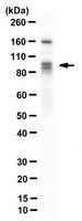
Evaluated by Western Blotting in HEK293 cell lysate.
Western Blotting Analysis: A 1:1,000 dilution of this antibody detected DYRK1A in 10 µg of HEK293 cell lysate.
Usage Statement
Unless otherwise stated in our catalog or other company documentation accompanying the product(s), our products are intended for research use only and are not to be used for any other purpose, which includes but is not limited to, unauthorized commercial uses, in vitro diagnostic uses, ex vivo or in vivo therapeutic uses or any type of consumption or application to humans or animals.