Wenn Sie das Fenster schließen, wird Ihre Konfiguration nicht gespeichert, es sei denn, Sie haben Ihren Artikel in die Bestellung aufgenommen oder zu Ihren Favoriten hinzugefügt.
Klicken Sie auf OK, um das MILLIPLEX® MAP-Tool zu schließen oder auf Abbrechen, um zu Ihrer Auswahl zurückzukehren.
Wählen Sie konfigurierbare Panels & Premixed-Kits - ODER - Kits für die zelluläre Signaltransduktion & MAPmates™
Konfigurieren Sie Ihre MILLIPLEX® MAP-Kits und lassen sich den Preis anzeigen.
Konfigurierbare Panels & Premixed-Kits
Unser breites Angebot enthält Multiplex-Panels, für die Sie die Analyten auswählen können, die am besten für Ihre Anwendung geeignet sind. Unter einem separaten Register können Sie das Premixed-Cytokin-Format oder ein Singleplex-Kit wählen.
Kits für die zelluläre Signaltransduktion & MAPmates™
Wählen Sie gebrauchsfertige Kits zur Erforschung gesamter Signalwege oder Prozesse. Oder konfigurieren Sie Ihre eigenen Kits mit Singleplex MAPmates™.
Die folgenden MAPmates™ sollten nicht zusammen analysiert werden: -MAPmates™, die einen unterschiedlichen Assaypuffer erfordern. -Phosphospezifische und MAPmate™ Gesamtkombinationen wie Gesamt-GSK3β und Gesamt-GSK3β (Ser 9). -PanTyr und locusspezifische MAPmates™, z.B. Phospho-EGF-Rezeptor und Phospho-STAT1 (Tyr701). -Mehr als 1 Phospho-MAPmate™ für ein einziges Target (Akt, STAT3). -GAPDH und β-Tubulin können nicht mit Kits oder MAPmates™, die panTyr enthalten, analysiert werden.
.
Bestellnummer
Bestellinformationen
St./Pkg.
Liste
Dieser Artikel wurde zu Ihren Favoriten hinzugefügt.
Wählen Sie bitte Spezies, Panelart, Kit oder Probenart
Um Ihr MILLIPLEX® MAP-Kit zu konfigurieren, wählen Sie zunächst eine Spezies, eine Panelart und/oder ein Kit.
Custom Premix Selecting "Custom Premix" option means that all of the beads you have chosen will be premixed in manufacturing before the kit is sent to you.
Catalogue Number
Ordering Description
Qty/Pack
List
Dieser Artikel wurde zu Ihren Favoriten hinzugefügt.
Spezies
Panelart
Gewähltes Kit
Menge
Bestellnummer
Bestellinformationen
St./Pkg.
Listenpreis
96-Well Plate
Menge
Bestellnummer
Bestellinformationen
St./Pkg.
Listenpreis
Weitere Reagenzien hinzufügen (MAPmates erfordern die Verwendung eines Puffer- und Detektionskits)
Menge
Bestellnummer
Bestellinformationen
St./Pkg.
Listenpreis
48-602MAG
Buffer Detection Kit for Magnetic Beads
1 Kit
Platzsparende Option Kunden, die mehrere Kits kaufen, können ihre Multiplex-Assaykomponenten in Kunststoffbeuteln anstelle von Packungen erhalten, um eine kompaktere Lagerung zu ermöglichen.
Dieser Artikel wurde zu Ihren Favoriten hinzugefügt.
Das Produkt wurde in Ihre Bestellung aufgenommen
Sie können nun ein weiteres Kit konfigurieren, ein Premixed-Kit wählen, zur Kasse gehen oder das Bestell-Tool schließen.
Detect DUX4 using this mouse monoclonal Anti-DUX4 Antibody, clone P4H2, Cat. No. MABS1348, validated for use in Immunocytochemistry and Western Blotting.
More>>Detect DUX4 using this mouse monoclonal Anti-DUX4 Antibody, clone P4H2, Cat. No. MABS1348, validated for use in Immunocytochemistry and Western Blotting. Less<<
Anti-DUX4 Antibody, clone P4H2: SDB (Sicherheitsdatenblätter), Analysenzertifikate und Qualitätszertifikate, Dossiers, Broschüren und andere verfügbare Dokumente.
Double homeobox protein 4 (UniProt Q9UBX2; also known as Double homeobox protein 10) is encoded by the DUX4 (also known as DUX10) gene (Gene ID 100288687) in human. DUX4 is a double homeobox transcription factor that is normally expressed in the testis. Contraction or hypomethylation of D4Z4 macrosatellite repeats on chromosome 4q35 causes aberrant DUX4 expression in skeletal muscle, a hallmark of the incurable disease facioscapulohumeral muscular dystrophy (FSHD), characterized by skeletal muscle weakness and wasting. Gene expression data analysis identifies β-catenin as the main coordinator of FSHD-associated perturbation of protein interaction signallings, including those of canonical Wnt, HIF1-α and TNF-α. In addition, DUX4 expression causes proteolytic degradation of UPF1, a central component of the nonsense-mediated decay (NMD) machinery, and accumulation NMD substrate RNAs including DUX4 mRNA, such that NMD downregulation by DUX4 expression stabilizes DUX4 mRNA through a double-negative feedback loop in FSHD muscle cells. FSHD muscle expresses a low abundance DUX4 mRNA that produces the full-length DUX4 protein. In contrast to control skeletal muscle and most other somatic tissues, full-length DUX4 transcript and protein is expressed at relatively abundant levels in human testis, most likely in the germ-line cells. Induced pluripotent (iPS) cells also express full-length DUX4 and differentiation of iPS cells to embryoid bodies suppresses expression of full-length DUX4, whereas expression of full-length DUX4 persists in differentiated FSHD iPS cells.
References
Product Information
Format
Purified
Presentation
Purified mouse IgG1 in buffer containing 0.1 M Tris-Glycine (pH 7.4), 150 mM NaCl with 0.05% sodium azide.
Detect DUX4 using this mouse monoclonal Anti-DUX4 Antibody, clone P4H2, Cat. No. MABS1348, validated for use in Immunocytochemistry and Western Blotting.
Key Applications
Immunocytochemistry
Western Blotting
Application Notes
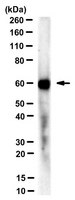
Western Blotting Analysis: 1 µg/mL from a representative lot detected DUX4 in 10 µg of human heart and ovary tissue lysates.
Immunocytochemistry Analysis: A representative lot detected full-length DUX4 (DUX4-FL) nuclear immunoreactivity at a higher frequency among differentiated CD56+ myogenic cells from facioscapulohumeral dystrophy (FSHD) than non-FSHD individuals (Jones, T.I., et al. (2012). Hum. Mol. Genet. 21(20):4419-4430).
Immunocytochemistry Analysis: A representative lot detected exogenously expressed full-length human DUX4 (DUX4-FL) in 2% paraformaldehyde-fixed and 1% Triton X-100-permeabilized C2C12 mouse myoblasts (Geng, L.N., et al. (2011). Hybridoma (Larchmt). 30(2):125-130).
Immunocytochemistry Analysis: A representative lot and another DUX4 antibody that targets an N-terminal region epitope co-stained full-length DUX4 (DUX4-FL) in the nucleus in ~0.1% of cultured facioscapulohumeral dystrophy (FSHD) muscle cells fixed with 2% paraformaldehyde and permeabilized with 1% Triton X-100 (Snider, L., et al. (2010). PLoS Genet. 6(10):e1001181).
Western Blotting Analysis: A representative lot detected exogenously expressed human DUX4, but not DUX4c, in transfected C2C12 mouse myoblasts. No target band was detected in lysate from untransfected cells (Geng, L.N., et al. (2011). Hybridoma (Larchmt). 30(2):125-130).
Western Blotting Analysis: A representative lot detected DUX4 immunoprecipitated from human testis tissue lysate by another DUX4 antibody that targets an N-terminal region epitope (Snider, L., et al. (2010). PLoS Genet. 6(10):e1001181).
Biological Information
Immunogen
GST-tagged recombinant human DUX4 C-terminal fragment.
Clone
P4H2
Concentration
Please refer to lot specific datasheet.
Host
Mouse
Specificity
Clone P4H2 is specific to DUX4 (UniProt Q9UBX2) and does not recognize DUX4c (DUX4L9; UniProt Q6RFH8) or the spliced DUX4 short isoform DUX4-s (UniProt E2JJS1) that lacks the C-terminal epitope region targeted by clone P4H2 (Geng, L.N., et al. (2011). Hybridoma (Larchmt). 30(2):125-130; Snider, L., et al. (2010). PLoS Genet. 6(10):e1001181).
~60/45 kDa observed. 44.94 kDa calculated. The larger-than-calculated target band size is consistent with that reported in the literature (Geng, L.N., et al. (2011). Hybridoma (Larchmt). 30(2):125-130; Snider, L., et al. (2010). PLoS Genet. 6(10):e1001181). Uncharacterized bands may be observed in some lysate(s).
Physicochemical Information
Dimensions
Materials Information
Toxicological Information
Safety Information according to GHS
Safety Information
Product Usage Statements
Quality Assurance
Evaluated by Western Blotting in human testis tissue lysate.
Western Blotting Analysis: 0.5 µg/mL of this antibody detected DUX4 in 10 µg of human testis tissue lysate.
Usage Statement
Unless otherwise stated in our catalog or other company documentation accompanying the product(s), our products are intended for research use only and are not to be used for any other purpose, which includes but is not limited to, unauthorized commercial uses, in vitro diagnostic uses, ex vivo or in vivo therapeutic uses or any type of consumption or application to humans or animals.