Wenn Sie das Fenster schließen, wird Ihre Konfiguration nicht gespeichert, es sei denn, Sie haben Ihren Artikel in die Bestellung aufgenommen oder zu Ihren Favoriten hinzugefügt.
Klicken Sie auf OK, um das MILLIPLEX® MAP-Tool zu schließen oder auf Abbrechen, um zu Ihrer Auswahl zurückzukehren.
Wählen Sie konfigurierbare Panels & Premixed-Kits - ODER - Kits für die zelluläre Signaltransduktion & MAPmates™
Konfigurieren Sie Ihre MILLIPLEX® MAP-Kits und lassen sich den Preis anzeigen.
Konfigurierbare Panels & Premixed-Kits
Unser breites Angebot enthält Multiplex-Panels, für die Sie die Analyten auswählen können, die am besten für Ihre Anwendung geeignet sind. Unter einem separaten Register können Sie das Premixed-Cytokin-Format oder ein Singleplex-Kit wählen.
Kits für die zelluläre Signaltransduktion & MAPmates™
Wählen Sie gebrauchsfertige Kits zur Erforschung gesamter Signalwege oder Prozesse. Oder konfigurieren Sie Ihre eigenen Kits mit Singleplex MAPmates™.
Die folgenden MAPmates™ sollten nicht zusammen analysiert werden: -MAPmates™, die einen unterschiedlichen Assaypuffer erfordern. -Phosphospezifische und MAPmate™ Gesamtkombinationen wie Gesamt-GSK3β und Gesamt-GSK3β (Ser 9). -PanTyr und locusspezifische MAPmates™, z.B. Phospho-EGF-Rezeptor und Phospho-STAT1 (Tyr701). -Mehr als 1 Phospho-MAPmate™ für ein einziges Target (Akt, STAT3). -GAPDH und β-Tubulin können nicht mit Kits oder MAPmates™, die panTyr enthalten, analysiert werden.
.
Bestellnummer
Bestellinformationen
St./Pkg.
Liste
Dieser Artikel wurde zu Ihren Favoriten hinzugefügt.
Wählen Sie bitte Spezies, Panelart, Kit oder Probenart
Um Ihr MILLIPLEX® MAP-Kit zu konfigurieren, wählen Sie zunächst eine Spezies, eine Panelart und/oder ein Kit.
Custom Premix Selecting "Custom Premix" option means that all of the beads you have chosen will be premixed in manufacturing before the kit is sent to you.
Catalogue Number
Ordering Description
Qty/Pack
List
Dieser Artikel wurde zu Ihren Favoriten hinzugefügt.
Spezies
Panelart
Gewähltes Kit
Menge
Bestellnummer
Bestellinformationen
St./Pkg.
Listenpreis
96-Well Plate
Menge
Bestellnummer
Bestellinformationen
St./Pkg.
Listenpreis
Weitere Reagenzien hinzufügen (MAPmates erfordern die Verwendung eines Puffer- und Detektionskits)
Menge
Bestellnummer
Bestellinformationen
St./Pkg.
Listenpreis
48-602MAG
Buffer Detection Kit for Magnetic Beads
1 Kit
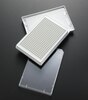
Platzsparende Option Kunden, die mehrere Kits kaufen, können ihre Multiplex-Assaykomponenten in Kunststoffbeuteln anstelle von Packungen erhalten, um eine kompaktere Lagerung zu ermöglichen.
Dieser Artikel wurde zu Ihren Favoriten hinzugefügt.
Das Produkt wurde in Ihre Bestellung aufgenommen
Sie können nun ein weiteres Kit konfigurieren, ein Premixed-Kit wählen, zur Kasse gehen oder das Bestell-Tool schließen.
AP1035
Sigma-AldrichAnti-Cyclophilin D Mouse mAb (E11AE12BD4)
This Anti-Cyclophilin D Mouse mAb (E11AE12BD4) is validated for use in Immunoblotting, Immunocytochemistry for the detection of Cyclophilin D.
More>>This Anti-Cyclophilin D Mouse mAb (E11AE12BD4) is validated for use in Immunoblotting, Immunocytochemistry for the detection of Cyclophilin D. Less<<
Anti-Cyclophilin D Mouse mAb (E11AE12BD4): SDB (Sicherheitsdatenblätter), Analysenzertifikate und Qualitätszertifikate, Dossiers, Broschüren und andere verfügbare Dokumente.
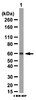
Recognizes the ~17-18 kDa processed form of mitochondrial cyclophilin D in cultured fibroblasts.
Catalogue Number
AP1035
Brand Family
Calbiochem®
Synonyms
Anti-CyPD
Application Data
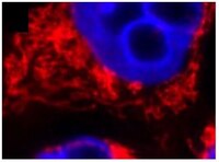
Detection of human cyclophilin D by immunoblotting. Sample: Heart mitochondria. Primary antibody: Anti-Cyclophilin D Mouse mAb (E11AE12BD4) (Cat. No. AP1035) (1 μg/ml). Detection: chemiluminescence. Detection of human cyclophilin D by immunocytochemistry. Sample: Cultured fibroblasts. Primary antibody: Anti-Cyclophilin D Mouse mAb (E11AE12BD4) (Cat. No. AP1035) (5 μg/ml). Detection: fluorescence.
References
References
Halestrap, A.P., et al. 2003. Curr. Med. Chem.10, 1507.
Anti-Cyclophilin D Mouse mAb (E11AE12BD4) Analysenzertifikate
Titel
Chargennummer
AP1035
Literatur
Übersicht
Halestrap, A.P., et al. 2003. Curr. Med. Chem.10, 1507.
Datenblatt
Note that this data sheet is not lot-specific and is representative of the current specifications for this product. Please consult the vial label and the certificate of analysis for information on specific lots. Also note that shipping conditions may differ from storage conditions.
Detection of human cyclophilin D by immunoblotting. Sample: Heart mitochondria. Primary antibody: Anti-Cyclophilin D Mouse mAb (E11AE12BD4) (Cat. No. AP1035) (1 μg/ml). Detection: chemiluminescence. Detection of human cyclophilin D by immunocytochemistry. Sample: Cultured fibroblasts. Primary antibody: Anti-Cyclophilin D Mouse mAb (E11AE12BD4) (Cat. No. AP1035) (5 μg/ml). Detection: fluorescence.
Description
Mouse monoclonal antibody purified by ammonium sulfate precipitation. Recognizes the ~17-18 kDa processed form of mitochondrial cyclophilin D (CyPD) protein.
Background
Cyclophilin D is a member of the immunophilin family of soluble cytosolic receptors capable of binding to one of two major immunosuppressant agents, cyclosporin A (CsA) or FK506. Proteins that bind CsA are called cyclophilins. Immunophilins function as peptidyl prolyl cis-trans-isomerases (PPlase) whose activity is inhibited by the respective immunosuppressant compounds. Immunophilins accelerate folding of proteins in vivo and in vitro by catalyzing slow steps in the initial folding and rearrangement of proline-containing proteins. Cyclophilin D is located in the intermembrane space of the mitochondria where it is an integral member of the permeability transition pore complex that includes the voltage dependent anion channel (VDAC) and adenine nucleotide translocase (ANT).
Host
Mouse
Immunogen species
Rat
Immunogen
recombinant, rat cyclophilin D
Clone
E11AE12BD4
Isotype
IgG₁
Species
bovine, human, mouse, rat
Positive control
Heart mitochondria
Form
Liquid
Formulation
In HBS.
Concentration Label
Please refer to vial label for lot-specific concentration
Preservative
≤0.1% sodium azide
Comments
Useful for detection of apoptosis related proteins. Antibody should be titrated for optimal results in individual systems.
Storage
Avoid freeze/thaw
-20°C
Do Not Freeze
Ok to freeze
Special Instructions
Following initial thaw, aliquot and freeze (-20°C).
Toxicity
Standard Handling
References
Halestrap, A.P., et al. 2003. Curr. Med. Chem.10, 1507.