Wenn Sie das Fenster schließen, wird Ihre Konfiguration nicht gespeichert, es sei denn, Sie haben Ihren Artikel in die Bestellung aufgenommen oder zu Ihren Favoriten hinzugefügt.
Klicken Sie auf OK, um das MILLIPLEX® MAP-Tool zu schließen oder auf Abbrechen, um zu Ihrer Auswahl zurückzukehren.
Wählen Sie konfigurierbare Panels & Premixed-Kits - ODER - Kits für die zelluläre Signaltransduktion & MAPmates™
Konfigurieren Sie Ihre MILLIPLEX® MAP-Kits und lassen sich den Preis anzeigen.
Konfigurierbare Panels & Premixed-Kits
Unser breites Angebot enthält Multiplex-Panels, für die Sie die Analyten auswählen können, die am besten für Ihre Anwendung geeignet sind. Unter einem separaten Register können Sie das Premixed-Cytokin-Format oder ein Singleplex-Kit wählen.
Kits für die zelluläre Signaltransduktion & MAPmates™
Wählen Sie gebrauchsfertige Kits zur Erforschung gesamter Signalwege oder Prozesse. Oder konfigurieren Sie Ihre eigenen Kits mit Singleplex MAPmates™.
Die folgenden MAPmates™ sollten nicht zusammen analysiert werden: -MAPmates™, die einen unterschiedlichen Assaypuffer erfordern. -Phosphospezifische und MAPmate™ Gesamtkombinationen wie Gesamt-GSK3β und Gesamt-GSK3β (Ser 9). -PanTyr und locusspezifische MAPmates™, z.B. Phospho-EGF-Rezeptor und Phospho-STAT1 (Tyr701). -Mehr als 1 Phospho-MAPmate™ für ein einziges Target (Akt, STAT3). -GAPDH und β-Tubulin können nicht mit Kits oder MAPmates™, die panTyr enthalten, analysiert werden.
.
Bestellnummer
Bestellinformationen
St./Pkg.
Liste
Dieser Artikel wurde zu Ihren Favoriten hinzugefügt.
Wählen Sie bitte Spezies, Panelart, Kit oder Probenart
Um Ihr MILLIPLEX® MAP-Kit zu konfigurieren, wählen Sie zunächst eine Spezies, eine Panelart und/oder ein Kit.
Custom Premix Selecting "Custom Premix" option means that all of the beads you have chosen will be premixed in manufacturing before the kit is sent to you.
Catalogue Number
Ordering Description
Qty/Pack
List
Dieser Artikel wurde zu Ihren Favoriten hinzugefügt.
Spezies
Panelart
Gewähltes Kit
Menge
Bestellnummer
Bestellinformationen
St./Pkg.
Listenpreis
96-Well Plate
Menge
Bestellnummer
Bestellinformationen
St./Pkg.
Listenpreis
Weitere Reagenzien hinzufügen (MAPmates erfordern die Verwendung eines Puffer- und Detektionskits)
Menge
Bestellnummer
Bestellinformationen
St./Pkg.
Listenpreis
48-602MAG
Buffer Detection Kit for Magnetic Beads
1 Kit
Platzsparende Option Kunden, die mehrere Kits kaufen, können ihre Multiplex-Assaykomponenten in Kunststoffbeuteln anstelle von Packungen erhalten, um eine kompaktere Lagerung zu ermöglichen.
Dieser Artikel wurde zu Ihren Favoriten hinzugefügt.
Das Produkt wurde in Ihre Bestellung aufgenommen
Sie können nun ein weiteres Kit konfigurieren, ein Premixed-Kit wählen, zur Kasse gehen oder das Bestell-Tool schließen.
Anti-CaV1.3 (809-825) Rabbit pAb: SDB (Sicherheitsdatenblätter), Analysenzertifikate und Qualitätszertifikate, Dossiers, Broschüren und andere verfügbare Dokumente.
Mitex Membran, PTFE, hydrophob, 5,0 µm, 142 mm, weiß, glatt
Übersicht
Replacement Information
Key Spec Table
Host
Rb
Description
Overview
This product has been discontinued.
Recognizes all forms of the α1D-subunit of Cav1.3 voltage-gated calcium channel. Supplied with a control peptide.
Note: The α-subunit is highly sensitive to proteases. Please refer to the product insert for proper sample preparation.
Catalogue Number
681509
Brand Family
Calbiochem®
Synonyms
Anti-L-Type Ca2+ Channel
Application Data
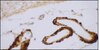
Detection of rat CaV1.3 by immunoblotting. Samples: Extract from rat brain membranes left untreated (lane 1) and treated by preincubation with the peptide antigen (lane 2). Primary antibody: Anti-CaV1.3 (809-825) Rabbit pAb (Cat. No. 681509) (1:200). Detection: chemilumiescence. Detection of rat CaV1.3 by immunohistochemistry. Samples: Adult rat dorsal root ganglion with staining appearing in clusters of cells (green) but not in axons. Staining of neurofilament 200 demonstrates partial overlap with CaV1.3 in neuronal staining but not in axons (red). Primary antibody: Anti-CaV1.3 (809-825) Rabbit pAb (Cat. No. 681509) (1:100). Detection: fluorescence.
References
References
Hell, J.W., et al. 1993. J. Cell Biol. 123, 949. Dubel, S.J., et al. 1992. Proc. Natl. Acad. Sci. USA 89, 5058. Hui, A., et al. 1991. Neuron7, 35.
Product Information
Form
Lyophilized
Formulation
50 µl antibody lyophilized from PBS, 5% sucrose, 1% BSA, 0.05% sodium azide, pH. 7.4 and 40 µg control peptide lyophilized from PBS.
The epitope is specific for α1D-subunits of the voltage-gated Ca2+ channel and is not present in any other known protein. It is highly conserved in human and hamster protein but cross-reactivity has not been tested. Antibody binding can be fully inhibited with the soluble CP809-825 blocking peptide (supplied). For use as a negative control, preincubate 1 µg control peptide with 1 µg antibody for 1 hr at room temperature. Caution: α1-subunits of voltage-gated Ca2+ channels are highly sensitive to proteases. All procedures involving a full-length protein should be performed at 4°C and include the following protease inhibitor cocktail: pepstatin A (Cat. No. 516482; 1 µg/ml), leupeptin (Cat. No. 108975; 1 µg/ml), aprotinin (Cat. No. 616398; 1 µg/ml), benzamidine (Cat. No. 199001; 0.1 µg/ml), AEBSF (Cat. No. 101500; 0.2 mM) and calpain inhibitors I (LLNL, Cat. No. 208719; 8 µg/ml) and II (ALLM, Cat. No. 208721; 8 µg/ml). To reduce nonspecific staining, centrifuge all antibody preparations before use (10000 x g for 5 min) to remove any aggregates. Variables associated with assay conditions will dictate the optimal working dilution.
Biological Information
Immunogen
a synthetic peptide [(KY)DNKVTIDDYQEEAEDKD] corresponding to amino acids 809-825 of the α1D-subunit of the rat brain voltage-gated calcium channels
Immunogen
Rat
Host
Rabbit
Isotype
IgG
Physicochemical Information
Dimensions
Materials Information
Toxicological Information
Safety Information according to GHS
Safety Information
Product Usage Statements
Storage and Shipping Information
Ship Code
Ambient Temperature Only
Toxicity
Standard Handling
Storage
-20°C
Do not freeze
Ok to freeze
Special Instructions
Reconstitute the antibody with 50 µl dH₂O. Further dilutions should be made in a physiological buffer containing a carrier protein such as 1-3% BSA. Reconstitute the peptide with 100 µl dH₂O, aliquot, and freeze (-20°C).
Hell, J.W., et al. 1993. J. Cell Biol. 123, 949. Dubel, S.J., et al. 1992. Proc. Natl. Acad. Sci. USA 89, 5058. Hui, A., et al. 1991. Neuron7, 35.
Datenblatt
Note that this data sheet is not lot-specific and is representative of the current specifications for this product. Please consult the vial label and the certificate of analysis for information on specific lots. Also note that shipping conditions may differ from storage conditions.
Detection of rat CaV1.3 by immunoblotting. Samples: Extract from rat brain membranes left untreated (lane 1) and treated by preincubation with the peptide antigen (lane 2). Primary antibody: Anti-CaV1.3 (809-825) Rabbit pAb (Cat. No. 681509) (1:200). Detection: chemilumiescence. Detection of rat CaV1.3 by immunohistochemistry. Samples: Adult rat dorsal root ganglion with staining appearing in clusters of cells (green) but not in axons. Staining of neurofilament 200 demonstrates partial overlap with CaV1.3 in neuronal staining but not in axons (red). Primary antibody: Anti-CaV1.3 (809-825) Rabbit pAb (Cat. No. 681509) (1:100). Detection: fluorescence.
Description
Immunoaffinity purified rabbit polyclonal antibody. Recognizes all forms of the α1D-subunit of the voltage-gated Ca channel. Supplied with a control peptide.
Host
Rabbit
Immunogen species
Rat
Immunogen
a synthetic peptide [(KY)DNKVTIDDYQEEAEDKD] corresponding to amino acids 809-825 of the α1D-subunit of the rat brain voltage-gated calcium channels
Isotype
IgG
Species
mouse, rat
Form
Lyophilized
Formulation
50 µl antibody lyophilized from PBS, 5% sucrose, 1% BSA, 0.05% sodium azide, pH. 7.4 and 40 µg control peptide lyophilized from PBS.
Preservative
≤0.1% sodium azide (antibody only)
Comments
The epitope is specific for α1D-subunits of the voltage-gated Ca2+ channel and is not present in any other known protein. It is highly conserved in human and hamster protein but cross-reactivity has not been tested. Antibody binding can be fully inhibited with the soluble CP809-825 blocking peptide (supplied). For use as a negative control, preincubate 1 µg control peptide with 1 µg antibody for 1 hr at room temperature. Caution: α1-subunits of voltage-gated Ca2+ channels are highly sensitive to proteases. All procedures involving a full-length protein should be performed at 4°C and include the following protease inhibitor cocktail: pepstatin A (Cat. No. 516482; 1 µg/ml), leupeptin (Cat. No. 108975; 1 µg/ml), aprotinin (Cat. No. 616398; 1 µg/ml), benzamidine (Cat. No. 199001; 0.1 µg/ml), AEBSF (Cat. No. 101500; 0.2 mM) and calpain inhibitors I (LLNL, Cat. No. 208719; 8 µg/ml) and II (ALLM, Cat. No. 208721; 8 µg/ml). To reduce nonspecific staining, centrifuge all antibody preparations before use (10000 x g for 5 min) to remove any aggregates. Variables associated with assay conditions will dictate the optimal working dilution.
Storage
-20°C
Do Not Freeze
Ok to freeze
Special Instructions
Reconstitute the antibody with 50 µl dH₂O. Further dilutions should be made in a physiological buffer containing a carrier protein such as 1-3% BSA. Reconstitute the peptide with 100 µl dH₂O, aliquot, and freeze (-20°C).
Toxicity
Standard Handling
References
Hell, J.W., et al. 1993. J. Cell Biol. 123, 949. Dubel, S.J., et al. 1992. Proc. Natl. Acad. Sci. USA 89, 5058. Hui, A., et al. 1991. Neuron7, 35.