Wenn Sie das Fenster schließen, wird Ihre Konfiguration nicht gespeichert, es sei denn, Sie haben Ihren Artikel in die Bestellung aufgenommen oder zu Ihren Favoriten hinzugefügt.
Klicken Sie auf OK, um das MILLIPLEX® MAP-Tool zu schließen oder auf Abbrechen, um zu Ihrer Auswahl zurückzukehren.
Wählen Sie konfigurierbare Panels & Premixed-Kits - ODER - Kits für die zelluläre Signaltransduktion & MAPmates™
Konfigurieren Sie Ihre MILLIPLEX® MAP-Kits und lassen sich den Preis anzeigen.
Konfigurierbare Panels & Premixed-Kits
Unser breites Angebot enthält Multiplex-Panels, für die Sie die Analyten auswählen können, die am besten für Ihre Anwendung geeignet sind. Unter einem separaten Register können Sie das Premixed-Cytokin-Format oder ein Singleplex-Kit wählen.
Kits für die zelluläre Signaltransduktion & MAPmates™
Wählen Sie gebrauchsfertige Kits zur Erforschung gesamter Signalwege oder Prozesse. Oder konfigurieren Sie Ihre eigenen Kits mit Singleplex MAPmates™.
Die folgenden MAPmates™ sollten nicht zusammen analysiert werden: -MAPmates™, die einen unterschiedlichen Assaypuffer erfordern. -Phosphospezifische und MAPmate™ Gesamtkombinationen wie Gesamt-GSK3β und Gesamt-GSK3β (Ser 9). -PanTyr und locusspezifische MAPmates™, z.B. Phospho-EGF-Rezeptor und Phospho-STAT1 (Tyr701). -Mehr als 1 Phospho-MAPmate™ für ein einziges Target (Akt, STAT3). -GAPDH und β-Tubulin können nicht mit Kits oder MAPmates™, die panTyr enthalten, analysiert werden.
.
Bestellnummer
Bestellinformationen
St./Pkg.
Liste
Dieser Artikel wurde zu Ihren Favoriten hinzugefügt.
Wählen Sie bitte Spezies, Panelart, Kit oder Probenart
Um Ihr MILLIPLEX® MAP-Kit zu konfigurieren, wählen Sie zunächst eine Spezies, eine Panelart und/oder ein Kit.
Custom Premix Selecting "Custom Premix" option means that all of the beads you have chosen will be premixed in manufacturing before the kit is sent to you.
Catalogue Number
Ordering Description
Qty/Pack
List
Dieser Artikel wurde zu Ihren Favoriten hinzugefügt.
Spezies
Panelart
Gewähltes Kit
Menge
Bestellnummer
Bestellinformationen
St./Pkg.
Listenpreis
96-Well Plate
Menge
Bestellnummer
Bestellinformationen
St./Pkg.
Listenpreis
Weitere Reagenzien hinzufügen (MAPmates erfordern die Verwendung eines Puffer- und Detektionskits)
Menge
Bestellnummer
Bestellinformationen
St./Pkg.
Listenpreis
48-602MAG
Buffer Detection Kit for Magnetic Beads
1 Kit
Platzsparende Option Kunden, die mehrere Kits kaufen, können ihre Multiplex-Assaykomponenten in Kunststoffbeuteln anstelle von Packungen erhalten, um eine kompaktere Lagerung zu ermöglichen.
Dieser Artikel wurde zu Ihren Favoriten hinzugefügt.
Das Produkt wurde in Ihre Bestellung aufgenommen
Sie können nun ein weiteres Kit konfigurieren, ein Premixed-Kit wählen, zur Kasse gehen oder das Bestell-Tool schließen.
ABE1957
Sigma-AldrichAnti-CENP-C Antibody
Anti-CENP-C Antibody, Cat. No. ABE1957, is a highly specific rabbit polyclonal antibody that targets CENP-C and has been tested in Immunocytochemistry and Western Blotting.
More>>Anti-CENP-C Antibody, Cat. No. ABE1957, is a highly specific rabbit polyclonal antibody that targets CENP-C and has been tested in Immunocytochemistry and Western Blotting. Less<<
Anti-CENP-C Antibody: SDB (Sicherheitsdatenblätter), Analysenzertifikate und Qualitätszertifikate, Dossiers, Broschüren und andere verfügbare Dokumente.
Centromere protein C (UniProt Q03188; also known as CENP-C, CENP-C 1, Centromere autoantigen C, Centromere protein C 1, Interphase centromere complex protein 7) is encoded by the CENPC (also known as CENPC1, ICEN7) gene (Gene ID 1060) in human. Kinetochores assemble on centromeres to bind spindle microtubules and mediate chromosome segregation during cell division. CENP-C is a subunit of constitutive centromere-associated network (CCAN) that acts as the centromere-kinetochore interface. CCAN is a large complex composed of at least 16 different centromeric proteins (CENPs). CENP-C plays an important role in kinetochore assembly downstream of CENP-A. The PEST domain in the N-terminal half of CENP-C interacts directly with the CCAN subcomplex CENP-HIKM (CENP-H, CENP-K, CENP-I, and CENP-M) and CENP-C depletion is shown to cause mislocalization of CENP-T and the CENP-HIKM complex subunits in HeLa cells.
References
Product Information
Format
Serum
Presentation
Rabbit polyclonal antibody serum with 0.05% sodium azide.
Anti-CENP-C Antibody, Cat. No. ABE1957, is a highly specific rabbit polyclonal antibody that targets CENP-C and has been tested in Immunocytochemistry and Western Blotting.
Key Applications
Western Blotting
Immunocytochemistry
Application Notes
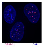
Immunocytochemistry Analysis: A 1:10,000 dilution from a representative lot immunostained kinetochores in chromosome spreads prepared from interphase DLD-1 human colorectal adenocarcinoma cells hypotonically swollen and fixed with 4% formaldehyde.
Immunocytochemistry Analysis: A representative lot was affinity purified and detected a time-dependent loss of kinetochores CENP-C immunoreactivity in 4% formaldehyde-fixed, 0.5% Triton X-100-permeabilized HeLa cells following CENP-C shRNA induction (Falk, S.J., et al. (2015). Science. 348(6235):699-703).
Immunocytochemistry Analysis: A representative lot was affinity purified and immunostained kinetochores by indirect fluorescence staining of chromosome spreads prepared from patient-derived, PD-NC4 chromosome variant harboring fibroblasts hypotonically swollen and fixed with 4% formaldehyde (Bassett, E.A., et al. (2010). J. Cell Biol. 190(2):177-185).
Western Blotting Analysis: A representative lot was affinity purified and detected a time-dependent CENP-C level in HeLa cells following CENP-C shRNA induction (Falk, S.J., et al. (2015). Science. 348(6235):699-703).
Biological Information
Immunogen
GST-tagged recombinant human CENP-C N-terminal fragment.
Epitope
N-terminus
Concentration
Please refer to lot specific datasheet.
Host
Rabbit
Specificity
This rabbit polyclonal antiserum targets the N-terminal region of human CENP-C present in both spliced isoforms reported by UniProt (Q03188). Specificity was demonstrated by a reducion of target band detection (Western blotting) and kinetochores staining (chromsome spreads fluoresence staining) following cellular CENP-C shRNA induction (Falk, S.J., et al. (2015). Science. 348(6235):699-703).
~140 kDa observed. 106.8 kDa (isoform 1) and 61.54 kDa (isoform 2) calculated. The larger-than-calculated target band size is consistent with that reported in the literature (Earnshaw, W.C., et al. (1989). Chromosoma. 98(1):1-12). Uncharacterized bands may be observed in some lysate(s).
Physicochemical Information
Dimensions
Materials Information
Toxicological Information
Safety Information according to GHS
Safety Information
Product Usage Statements
Quality Assurance
Evaluated by Western Blotting in DLD-1 cell lysate.
Western Blotting Analysis: A 1:5,000 dilution of this antiserum detected CENP-C in 50,000 cell equivalent of DLD-1 human colorectal adenocarcinoma cell lysate.
Usage Statement
Unless otherwise stated in our catalog or other company documentation accompanying the product(s), our products are intended for research use only and are not to be used for any other purpose, which includes but is not limited to, unauthorized commercial uses, in vitro diagnostic uses, ex vivo or in vivo therapeutic uses or any type of consumption or application to humans or animals.
Storage and Shipping Information
Storage Conditions
Stable for 1 year at -20°C from date of receipt. Handling Recommendations: Upon receipt and prior to removing the cap, centrifuge the vial and gently mix the solution. Aliquot into microcentrifuge tubes and store at -20°C. Avoid repeated freeze/thaw cycles, which may damage IgG and affect product performance.