Wenn Sie das Fenster schließen, wird Ihre Konfiguration nicht gespeichert, es sei denn, Sie haben Ihren Artikel in die Bestellung aufgenommen oder zu Ihren Favoriten hinzugefügt.
Klicken Sie auf OK, um das MILLIPLEX® MAP-Tool zu schließen oder auf Abbrechen, um zu Ihrer Auswahl zurückzukehren.
Wählen Sie konfigurierbare Panels & Premixed-Kits - ODER - Kits für die zelluläre Signaltransduktion & MAPmates™
Konfigurieren Sie Ihre MILLIPLEX® MAP-Kits und lassen sich den Preis anzeigen.
Konfigurierbare Panels & Premixed-Kits
Unser breites Angebot enthält Multiplex-Panels, für die Sie die Analyten auswählen können, die am besten für Ihre Anwendung geeignet sind. Unter einem separaten Register können Sie das Premixed-Cytokin-Format oder ein Singleplex-Kit wählen.
Kits für die zelluläre Signaltransduktion & MAPmates™
Wählen Sie gebrauchsfertige Kits zur Erforschung gesamter Signalwege oder Prozesse. Oder konfigurieren Sie Ihre eigenen Kits mit Singleplex MAPmates™.
Die folgenden MAPmates™ sollten nicht zusammen analysiert werden: -MAPmates™, die einen unterschiedlichen Assaypuffer erfordern. -Phosphospezifische und MAPmate™ Gesamtkombinationen wie Gesamt-GSK3β und Gesamt-GSK3β (Ser 9). -PanTyr und locusspezifische MAPmates™, z.B. Phospho-EGF-Rezeptor und Phospho-STAT1 (Tyr701). -Mehr als 1 Phospho-MAPmate™ für ein einziges Target (Akt, STAT3). -GAPDH und β-Tubulin können nicht mit Kits oder MAPmates™, die panTyr enthalten, analysiert werden.
.
Bestellnummer
Bestellinformationen
St./Pkg.
Liste
Dieser Artikel wurde zu Ihren Favoriten hinzugefügt.
Wählen Sie bitte Spezies, Panelart, Kit oder Probenart
Um Ihr MILLIPLEX® MAP-Kit zu konfigurieren, wählen Sie zunächst eine Spezies, eine Panelart und/oder ein Kit.
Custom Premix Selecting "Custom Premix" option means that all of the beads you have chosen will be premixed in manufacturing before the kit is sent to you.
Catalogue Number
Ordering Description
Qty/Pack
List
Dieser Artikel wurde zu Ihren Favoriten hinzugefügt.
Spezies
Panelart
Gewähltes Kit
Menge
Bestellnummer
Bestellinformationen
St./Pkg.
Listenpreis
96-Well Plate
Menge
Bestellnummer
Bestellinformationen
St./Pkg.
Listenpreis
Weitere Reagenzien hinzufügen (MAPmates erfordern die Verwendung eines Puffer- und Detektionskits)
Menge
Bestellnummer
Bestellinformationen
St./Pkg.
Listenpreis
48-602MAG
Buffer Detection Kit for Magnetic Beads
1 Kit
Platzsparende Option Kunden, die mehrere Kits kaufen, können ihre Multiplex-Assaykomponenten in Kunststoffbeuteln anstelle von Packungen erhalten, um eine kompaktere Lagerung zu ermöglichen.
Dieser Artikel wurde zu Ihren Favoriten hinzugefügt.
Das Produkt wurde in Ihre Bestellung aufgenommen
Sie können nun ein weiteres Kit konfigurieren, ein Premixed-Kit wählen, zur Kasse gehen oder das Bestell-Tool schließen.
Detect Annexin A3 using this mouse monoclonal Anti-Annexin A3 Antibody, clone 9996-1-3M1/3L7-5, Cat. No. MABS1329, validated for use in Immunohistochemistry (Paraffin) Neutralization, and Western Blotting.
More>>Detect Annexin A3 using this mouse monoclonal Anti-Annexin A3 Antibody, clone 9996-1-3M1/3L7-5, Cat. No. MABS1329, validated for use in Immunohistochemistry (Paraffin) Neutralization, and Western Blotting. Less<<
SDB (Sicherheitsdatenblätter), Analysenzertifikate und Qualitätszertifikate, Dossiers, Broschüren und andere verfügbare Dokumente.
Annexin A3 (UniProt P12429; also known as 35-alpha calcimedin, Annexin III, Annexin-3, Inositol 1,2-cyclic phosphate 2-phosphohydrolase, Lipocortin III, PAP-III, Placental anticoagulant protein III) is encoded by the ANXA3 (also known as ANX3) gene (Gene ID 306) in human. Annexin A3 is a Ca2+-dependent phospholipid-binding protein shown to promote angiogenesis and rat liver regeneration. Upregulated annexin A3 expression is reported in ovarian, breast, colon, lung, gastric, gallbladder, testicular, and urothelial cancers, where it is involved in promoting tumorigenesis and resistance to chemotherapy. ANXA3 is reported to represent the most significantly upregulated gene that encodes for a secretory protein in the CD133+ liver cancer stem cell (CSC) subset. Both cellular and secretory annexin A3 play pivotal roles in promoting cancer and stem cell-like features in CD133+ liver CSCs through a dysregulated JNK pathway. Annexin A3 serum levels in hepatocellular carcinoma (HCC) patients are closely associated with aggressive clinical features. Annexin A3 neutralizing antibody is shown to cause a significant reduction in HCC tumor growth and self-renewal by effectively killing off CD133+ CSCs both in cultures in vitro and in animal xenograft in vivo.
References
Product Information
Format
Purified
Presentation
Purified mouse IgG1 in Tris-buffered saline (TBS) containing 50 mM tris-HCl (pH7.5) and 150 mM NaCl without preservatives.
Detect Annexin A3 using this mouse monoclonal Anti-Annexin A3 Antibody, clone 9996-1-3M1/3L7-5, Cat. No. MABS1329, validated for use in Immunohistochemistry (Paraffin) Neutralization, and Western Blotting.
Key Applications
Western Blotting
Immunohistochemistry (Paraffin)
Neutralization Assay
Application Notes
Immunohistochemistry Analysis: A 1:250 dilution from a representative lot detected Annexin A3 in human placenta tissue sections.
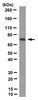
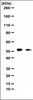
Western Blotting Analysis: 10 µg/mL from a representative lot detected Annexin A3 in 10 µg of human spleen tissue lysate.
Immunohistochemistry Analysis: A representative lot detected a markedly downregulated annexin A3 immunoreactivity in Huh7 xenografts from antibody-treated mice, while an upregulated annexin A3 immunoreactivity was seen in xenografts from Cisplatin-treated mice (Tong, M., et al. (2015). Stem Cell Reports. 5(1):45-59).
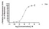
Neutralizing Analysis: A representative lot inhibited the proliferation of CD133-/annexin A3-positive Huh7 cells, but not the CD133-/annexin A3-negative MIHA cells. Antibody treatment further sensitized Huh7 to Cisplatin (Cat. No. 232120) antiproliferative activity (Tong, M., et al. (2015). Stem Cell Reports. 5(1):45-59).
Neutralizing Analysis: A representative lot suppressed the migration, invasion, angiogenesis induction, and hepatospheres formation of human hepatocellular carcinoma (HCC) cells (Tong, M., et al. (2015). Stem Cell Reports. 5(1):45-59).
Neutralizing Analysis: A representative lot inhibited the growth of Huh7 xenograph in mice. Residual xenografts following antibody treatment (alone or with Cisplatin), but not Cisplatin treatment alone failed to form tumor mass when serially transplanted into secondary recipients (Tong, M., et al. (2015). Stem Cell Reports. 5(1):45-59).
Western Blotting Analysis: A representative lot detected the expression of the ~33 kDa Annexin A3 among a panel of human hepatocellular carcinoma (HCC) cell lines (Tong, M., et al. (2015). Stem Cell Reports. 5(1):45-59).
Biological Information
Immunogen
Recombinant human annexin A3.
Clone
9996-1-3M1/3L7-5
Concentration
Please refer to lot specific datasheet.
Host
Mouse
Specificity
This monoclonal antibody detected a single ~33 kDa target band among a panel of human hepatocellular carcinoma (HCC) cell lines. The epitope has been mapped to a 10-a.a. region within the third annexin domain (Tong, M., et al. (2015). Stem Cell Reports. 5(1):45-59).
~33 kDa observed. 36.24 kDa calculated (Met1 removed). Uncharacterized bands may be observed in some lysate(s).
Physicochemical Information
Dimensions
Materials Information
Toxicological Information
Safety Information according to GHS
Safety Information
Product Usage Statements
Quality Assurance
Evaluated by Western Blotting in human placenta tissue lysate.
Western Blotting Analysis: 5 µg/mL of this antibody detected Annexin A3 in 10 µg of human placenta tissue lysate.
Usage Statement
Unless otherwise stated in our catalog or other company documentation accompanying the product(s), our products are intended for research use only and are not to be used for any other purpose, which includes but is not limited to, unauthorized commercial uses, in vitro diagnostic uses, ex vivo or in vivo therapeutic uses or any type of consumption or application to humans or animals.
Storage and Shipping Information
Storage Conditions
Stable for 1 year at -20°C from date of receipt. Handling Recommendations: Upon receipt and prior to removing the cap, centrifuge the vial and gently mix the solution. Aliquot into microcentrifuge tubes and store at -20°C. Avoid repeated freeze/thaw cycles, which may damage IgG and affect product performance.