Millipore Sigma Vibrant Logo

17-10230 LentiBrite™ GFP-LC3-II Enrichment Kit (Flow Cytometry)

17-10230
Minimum of 3 x 10E8 infectious units (IFU) per mL  
가격 검색...
가격을 검색할 수 없습니다
Minimum Quantity needs to be mulitiple of
Maximum Quantity is
가격 문의 추가 정보
()이 할인됨
 
가격 문의
현재 재고 없음
현재 재고 없음
예상 출고 가능일 
단종품
제한된 수량 가능
재고여부 확인
    Remaining: will advise
      Remaining: will advise
      추천사항
      고객 서비스로 문의
      Contact Customer Service


      고객 서비스로 문의

      개요

      Replacement Information
      Description
      Catalogue Number17-10230
      Trade Name
      • LentiBrite
      • Chemicon
      DescriptionLentiBrite™ GFP-LC3-II Enrichment Kit (Flow Cytometry)
      OverviewAutophagy, a degradative pathway that provides recycled nutrients to cells under stress, plays both protective and deleterious roles in many diseases, including cancer, neurodegeneration, and infections. Members of the LC3 family play a key role in the maturation of the autophagosome, the central organelle of autophagy. LC3 precursors, diffusely distributed in the cytosol, are proteolytically processed to form LC3-I. Upon initiation of autophagy, the C-terminal glycine is modified by addition of a phosphatidylethanolamine (PE) to form LC3-II, which translocates rapidly to nascent autophagosomes in a punctate distribution. DNA constructs encoding fluorescent proteins fused to LC3 are widely employed for introduction into cells for monitoring autophagosome formation by fluorescence microscopy.
      EMD Millipore’s LentiBrite™ GFP-LC3 lentiviral particles provide bright fluorescence and precise localization to enable live cell analysis of autophagy in difficult-to-transfect cell types. The

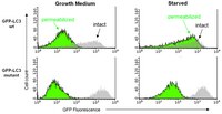
      LentiBrite™ GFP-LC3-II Enrichment Kit (Flow Cytometry) combines our GFP-LC3 lentiviral particles with a selective permeabilization procedure for GFP-LC3 transduced cells that allows release of cytosolic LC3-I and retention of autophagosome-bound LC3-II. This procedure allows detection of autophagosome density by flow cytometry.
      Materials Required but Not Delivered1. Test tubes for sample preparation, centrifugation and storage
      2. Tissue culture reagents, i.e. HBSS, PBS w/o Ca2+ or Mg2+, cell dislodging buffers, etc.
      3. Pipettors with corresponding tips capable of accurately measuring 10 – 1000 μL
      4. V-bottom 96-well plates (Corning 2897)
      5. Tabletop centrifuge capable of achieving 300 x g and holding 96-well plates
      6. Mechanical vortex
      7. Flow Cytometer
      8. Deionized water (for reagent dilutions)
      References
      Product Information
      Components
      • 1. TagGFP2-LC3 Lentivirus: One vial containing 25 µL of lentiviral particles at a minimum of 3 x 10E8 infectious units (IFU) per mL. For lot-specific titer information, please see datasheet.
        • Virus Description: 3rd generation HIV-based VSV-G pseudotyped lentiviral particles. Lentiviral particles were generated using the pPACKH1 Lentivector Packaging System and purified by PEG precipitation.
        • Promoter: EF-1 (Elongation Factor-1).
        • Expression Marker Description: TagGFP2 (Evrogen) fused at its C-terminus with human LC3A (Accession # NP_115903) truncated to leave G120 (lipidation site) at the C-terminus.
        (Store at -80°C)

        2. Autophagy Reagent A: One vial (lyophilized) (Store at 2-8°C)
        3. 10x Enrichment Buffer (Part No. CS210552): One vial containing 1 mL (Store at 2-8°C)
        4. 5x Assay Buffer: (Part No. CS210529): One bottle containing 3 mL (Store at 2-8°C)
      Detection methodFluorescent
      Quality LevelMQ100
      Applications
      Key Applications
      • Flow Cytometry
      Application NotesEMD Millipore’s LentiBrite™ GFP-LC3 lentiviral particles provide bright fluorescence and precise localization to enable live cell analysis of autophagy in difficult-to-transfect cell types. The LentiBrite™ GFP-LC3-II Enrichment Kit (Flow Cytometry) combines our GFP-LC3 lentiviral particles with a selective permeabilization procedure for GFP-LC3 transduced cells that allows release of cytosolic LC3-I and retention of autophagosome-bound LC3-II. This procedure allows detection of autophagosome density by flow cytometry.

      For optimal fluorescent analysis, it is recommended to analyze the target expression level within 24-48 hrs after transfection/infection for optimal live cell analysis, as fluorescent intensity may dim over time, especially in difficult-to-transfect cell lines. Infected cells may be frozen down after successful transfection/infection and thawed in culture to retain positive fluorescent expression beyond 24-48 hrs. Length and intensity of fluorescent expression varies between cell lines. Higher MOIs may be required for difficult-to-transfect cell lines.
      Biological Information
      Physicochemical Information
      Dimensions
      Materials Information
      Toxicological Information
      Safety Information according to GHS
      Safety Information
      Product Usage Statements
      Usage Statement
      • This product contains genetically modified organisms (GMO). Within the EU GMOs are regulated by Directives 2001/18/EC and 2009/41/EC of the European Parliament and of the Council and their national implementation in the member States respectively. This legislation obliges Merck to request certain information about you and the establishment where the GMOs are being handled. Click here for Enduser Declaration (EUD) Form.
      • Unless otherwise stated in our catalog or other company documentation accompanying the product(s), our products are intended for research use only and are not to be used for any other purpose, which includes but is not limited to, unauthorized commercial uses, in vitro diagnostic uses, ex vivo or in vivo therapeutic uses or any type of consumption or application to humans or animals.
      Storage and Shipping Information
      Storage ConditionsStore TagGFP2-LC3 Lentivirus at -80°C. After first thaw, place immediately on ice and freeze in working aliquots at -80°C. Frozen aliquots may be stored for at least 2 months. Further freeze/thaws may result in decreased virus titer and transduction efficiency.

      Store Autophagy Reagent A, 10x Enrichment Buffer, and 5x Assay Buffer
      at 2-8°C.

      All components are stable for at least 4 months from date of receipt when stored as recommended above.
      Packaging Information
      Material SizeMinimum of 3 x 10E8 infectious units (IFU) per mL
      Transport Information
      Supplemental Information
      Specifications
      Global Trade Item Number
      카탈로그 번호 GTIN
      17-10230 04053252008795

      Documentation

      LentiBrite™ GFP-LC3-II Enrichment Kit (Flow Cytometry) MSDS

      타이틀

      물질안전보건자료(MSDS) 

      LentiBrite™ GFP-LC3-II Enrichment Kit (Flow Cytometry) Certificates of Analysis

      TitleLot Number
      LentiBrite GFP-LC3-II Enrichment Kit (Flow Cytometry) - 2234231 2234231
      LentiBrite GFP-LC3-II Enrichment Kit (Flow Cytometry) - Q2007837 Q2007837
      LentiBrite™ GFP-LC3-II Enrichment Kit (Flow Cytometry) 3077700
      LentiBrite™ GFP-LC3-II Enrichment Kit (Flow Cytometry) - 3282694 3282694
      LentiBrite™ GFP-LC3-II Enrichment Kit (Flow Cytometry) - 3425324 3425324
      LentiBrite™ GFP-LC3-II Enrichment Kit (Flow Cytometry) - 3588666 3588666
      LentiBrite™ GFP-LC3-II Enrichment Kit (Flow Cytometry) -2562401 2562401
      LentiBrite™ GFP-LC3-II Enrichment Kit (Flow Cytometry) -2698470 2698470
      LentiBrite™ GFP-LC3-II Enrichment Kit (Flow Cytometry) - 2424153 2424153

      Technical Info

      Title
      LentiBrite™ Lentiviral Biosensors for Fluorescent Cellular Imaging: Analysis of Autophagosome Formation

      Posters

      Title
      Autophagy Signaling

      Newsletters / Publications

      Title
      Cellutions - The Newsletter for Cell Biology Researchers Vol 3:2012

      관련 제품 및 어플리케이션

      관련 제품: Brand Facete

      카테고리

      Life Science Research > Cell Analysis > guava easyCyte Flow Cytometers > Flow Cytometry Kits & Reagents