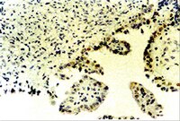
Differential expression of two new members of the p53 family, p63 and p73, in extramammary Paget's disease.
S Chen, Y Moroi, K Urabe, S Takeuchi, M Kido, S Hayashida, H Uchi, T Uenotsuchi, Y Tu, M Furue
Clinical and experimental dermatology
33
634-40
2008
요약 표시
BACKGROUND: The proteins p53, p63 and p73 are known to be overexpressed and to play important roles in the pathogenesis of many tumours, but the expression of p63 and p73 has not previously been investigated in extramammary Paget's disease (EMPD). AIM: To investigate the potential contribution of p53, p63 and p73 in the pathogenesis of EMPD. METHODS: In total, 35 paraffin wax-embedded tissue samples from patients with EMPD were examined using immunohistochemical staining for p53, p63 and p73. RESULTS: All of the 35 EMPD specimens, including all 6 invasive EMPD and 2 metastatic lymph-node specimens, showed nuclear overexpression of both p53 and p73. The expression levels (percentage of positive cells) of p53 and p73 (90.66 +/- 12.53% and 80.20 +/- 13.07%) in EMPD were significantly higher than those of normal skin. There was a significant correlation between the expression levels of p53 and p73 in EMPD. In 29 of 35 EMPD specimens, there was no nuclear expression of p63, and weak or moderate staining was found in only 6 specimens. The expression level of p63 in EMPD was significantly less than that in normal skin. CONCLUSIONS: Our study shows that the concordant overexpression of p53 and p73 and the decreased expression of p63 may play a pivotal role in the pathogenesis of EMPD. The decreased expression of p63 may play a more important role in the pathogenesis of EMPD than the overexpression of p53 and p73. | 18627398
 |
Overexpression of phosphorylated-ATF2 and STAT3 in cutaneous angiosarcoma and pyogenic granuloma.
Si-Yuan Chen,Satoshi Takeuchi,Kazunori Urabe,Sayaka Hayashida,Makiko Kido,Hiroto Tomoeda,Hiroshi Uchi,Teruki Dainichi,Masakazu Takahara,Satoko Shibata,Ya-Ting Tu,Masutaka Furue,Yoichi Moroi
Journal of cutaneous pathology
35
2008
요약 표시
Activating transcription factor-2/Activator protein-1 (AP-1), Signal transducer and activator of transcription-3 and p53 are important regulators of cellular proliferation, apoptosis, differentiation in the pathogenesis of many human tumors, but the expression of phosphorylated (p)-activating transcription factor-2 (p-ATF2), phosphorylated (p)-signal transducer and activator of transcription-3 (p-STAT3) and p53 family (p63 and p73) has not been investigated in cutaneous angiosarcoma (CAS) and pyogenic granuloma (PG) so far. | 18700251
 |
Expression of p73 in normal skin and proliferative skin lesions.
Makoto Kamiya, Yuko Takeuchi, Mari Katho, Hideaki Yokoo, Atsushi Sasaki, Yoichi Nakazato
Pathology international
54
890-5
2004
요약 표시
The p73 gene is a member of the p53 gene family and the structure and functions of p73 protein are similar to those of p53. However, these two proteins have different roles. In the present study, p73 protein was found immunohistochemically to be distributed in the basal cells of the epidermis, columnar basal cells in the hair follicle and peripheral cells without lipid droplets in the sebaceous and meibomian glands; it was expressed strongly in tumor cells in basal cell carcinomas and in the basal cell-like cells in seborrheic keratosis, and weakly or negatively in the squamous cell-like cells in seborrheic keratosis and in the tumor cells in squamous cell carcinomas. No relationship was detected between p73 and p53 protein distribution and between p73 protein expression and the proliferative potential, as shown by the Ki-67 immunopositive cell ratio. The present study shows that p73 protein is likely to play important roles in skin differentiation rather than proliferation or carcinogenesis of the skin. | 15598310
 |
The expression of p73, p21 and MDM2 proteins in gliomas.
Makoto Kamiya, Yoichi Nakazato, Makoto Kamiya, Yoichi Nakazato
Journal of neuro-oncology
59
143-9
2002
요약 표시
p73 protein, a member of the p53 family protein, induce p21 and MDM2 transcription. In some carcinomas, the expression of p73 was higher in carcinoma than in normal tissue, and the overexpression was correlated to grade, stage or prognosis. However, either the expression of p73 protein or the relationship among p73, p21 and MDM2 proteins was not known well in glioma. In this study, we examined the expression of p73, p21 and MDM2 proteins immunohistochemically and analyzed the relationship among these three proteins in sixty surgical specimens of gliomas including 10 glioblastomas, 10 anaplastic astrocytomas, 6 diffuse astrocytomas, 8 pilocytic astrocytomas, 1 anaplastic ependymoma, 8 ependymomas, 9 anaplastic oligodendroglial tumors and 8 low grade oligodendroglial tumors. The p73 labelling index (LI)s and p21 LIs differed among the tumor types, but there was no difference in the MDM2 LIs among all of the tumor types. The mean p73 LI of ependymomas was significantly higher than in any other tumor type. The mean p2I LIs of ependymomas and pilocytic astrocytomas were significantly higher than in any other tumor type. There were no significant correlations among p73 LIs, p21 LIs, MDM2 LIs and MIB-1 LIs in all p73 immunopositive tumors. The present results suggest that p73 and p21 overexpression of ependymomas and p21 overexpression of pilocytic astrocytomas are one of the features of these tumors, and that p73 overexpression does not influence the expression manners of either p21 or MDM2 in gliomas. | 12241107
 |
Differential expression of p53 gene family members p63 and p73 in head and neck squamous tumorigenesis.
Hong-Ran Choi, John G Batsakis, Feng Zhan, Erich Sturgis, Mario A Luna, Adel K El-Naggar
Human pathology
33
158-64
2002
요약 표시
p73 and p63 are recently cloned genes that share considerable structural and functional homologies with the p53 tumor suppressor gene. These genes, unlike p53, express multiple mRNA isoforms with variable biologic functions, and their suppressor nature has yet to be confirmed. To determine the interrelationship between these genes in the tumorigenesis of head and neck squamous carcinoma (HNSC), we performed immunohistochemical analyses of their protein products and compared the data with clinicopathologic parameters in 38 patients. In histologically normal epithelium, p53 and p73 showed similar basal and/or parabasal expression, but that of p53 was weaker and discontinuous. p63 staining was noted in more suprabasal cellular layers and was stronger. In dysplasias, all three markers manifested variable but gradual increase in extent and intensity of cellular expression with histologic progression. In carcinomas, p63 was the most frequently expressed (94.7%), followed by p73 (68.4%) and p53 (52.6%). Significant statistical correlation was noted only between p63 and p73 expressions (P =.04). Although no statistical correlation was found between p53 and p63 or p73, p53-negative tumors overexpressed either p63 or p73. p73 expression was associated with distant metastasis and perineural/vascular invasion. Our study indicates that (1) p63 and p73 expression may represent an early event in HNSC tumorigenesis, (2) the lack of correlation between p73 or p63 and p53 expression suggests an independent and/or compensatory functional role, (3) p73 expression may play a part in HNSC progression, and (4) p73 and p63 may function as oncogenes in the development of these tumors. | 11957139
 |
Monoallelically expressed gene related to p53 at 1p36, a region frequently deleted in neuroblastoma and other human cancers.
Kaghad, M, et al.
Cell, 90: 809-19 (1997)
1997
요약 표시
We describe a gene encoding p73, a protein that shares considerable homology with the tumor suppressor p53. p73 maps to 1p36, a region frequently deleted in neuroblastoma and other tumors and thought to contain multiple tumor suppressor genes. Our analysis of neuroblastoma cell lines with 1p and p73 loss of heterozygosity failed to detect coding sequence mutations in remaining p73 alleles. However, the demonstration that p73 is monoallelically expressed supports the notion that it is a candidate gene in neuroblastoma. p73 also has the potential to activate p53 target genes and to interact with p53. We propose that the disregulation of p73 contributes to tumorigenesis and that p53-related proteins operate in a network of developmental and cell cycle controls. | 9288759
 |
p73 is a simian [correction of human] p53-related protein that can induce apoptosis.
Jost, C A, et al.
Nature, 389: 191-4 (1997)
1997
요약 표시
The protein p53 is the most frequently mutated tumour suppressor to be identified so far in human cancers. The ability of p53 to inhibit cell growth is due, at least in part, to its ability to bind to specific DNA sequences and activate the transcription of target genes such as that encoding the cell-cycle inhibitor p21Waf1/Cip1 . A gene has recently been identified that is predicted to encode a protein with significant amino-acid sequence similarity to p53. In particular, each of the p53 amino-acid residues implicated in direct sequence-specific DNA binding is conserved in this protein. This gene, called p73, maps to the short arm of chromosome 1, and is found in a region that is frequently deleted in neuroblastomas. Here we show that p73 can, at least when overproduced, activate the transcription of p53-responsive genes and inhibit cell growth in a p53-like manner by inducing apoptosis (programmed cell death). | 9296498
 |


