Millipore Sigma Vibrant Logo

NR07 Anti-Ryanodine Receptor Mouse mAb (C3-33)

View Products on Sigmaaldrich.com
NR07
가격 및 재고 조회

개요

Replacement Information
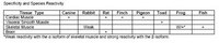
주요 사양표

Species ReactivityHostAntibody Type
Am, Av, Ca, F, Rb, RMMonoclonal Antibody

가격 및 재고여부

카탈로그 번호 재고 정보패킹 포장 단위 가격(VAT 별도) 수량
NR07-100UGCN
재고 정보 검색...
현재 재고 없음
현재 재고 없음
예상 출고 가능일 
단종품
제한된 수량 가능
재고여부 확인
    Remaining: will advise
      Remaining: will advise
      추천사항
      고객 서비스로 문의
      Contact Customer Service

      Plastic ampoule 100 μg
      가격 검색...
      가격을 검색할 수 없습니다
      Minimum Quantity needs to be mulitiple of
      Maximum Quantity is
      가격 문의 추가 정보
      ()이 할인됨
       
      가격 문의
      Description
      OverviewRecognizes the ~565 kDa ryanodine receptor in rat brain hippocampus. A ~565 kDa doublet representing the α and β isoforms is observed in non-mammalian vertebrates.
      Catalogue NumberNR07
      Brand Family Calbiochem®
      Application Data

      Detection of rat ryanodine receptor by immunocytochemistry. Sample: Rat brain hippocampus. Primary antibody: Anti-Ryanodine Receptor Mouse mAb (C3-33) (Cat. No. NR07) (1:50). Detection: fluorescence.
      References
      ReferencesSitsapesan, R., et al. 1995. Trends Pharmacol. Sci. 16, 386.
      McPherson, P.S. and Campbell, K.P. 1993. J. Biol. Chem. 268, 13765.
      Meszaros, L.G., et al. 1993. Nature 364, 76.
      Lai, F.A., et al. 1992. Biochem. J. 288, 553.
      Lai, F.A., et al. 1989. Nature 331, 315.
      Product Information
      FormLiquid
      FormulationIn PBS.
      Positive controlRat brain hippocampus
      Preservative≤0.1% sodium azide
      Quality LevelMQ100
      Applications
      Application ReferencesOriginal Clone Lai, F.A., et al. 1992. Amer. J. Physiol. 263, C365. Frozen Sections Junker, J., et al. 1994. J. Biol. Chem. 269, 1627.
      Key Applications Frozen Sections
      Immunoblotting (Western Blotting)
      Immunoprecipitation
      Application NotesFrozen Sections (1 µg/ml, see application references)
      Immunoblotting (1 µg/ml)
      Immunoprecipitation (see comments)
      Application CommentsA ~565 kDa doublet representing the α and β isoforms is observed in non-mammalian vertebrates. In frozen sections, Clone C3-33 strongly stains the myofiber consistent with the sarcoplasmic reticulum of the RyR. This antibody has been reported to work for immunoprecipitation; the concentration is assay-dependent. Antibody should be titrated for optimal results in individual systems.
      Biological Information
      Immunogencanine cardiac ryanodine receptor (RYR)
      ImmunogenCanine
      CloneC3-33
      HostMouse
      IsotypeIgG₁
      Species Reactivity
      • Amphibian
      • Avian
      • Canine
      • Fish
      • Rabbit
      • Rat
      Antibody TypeMonoclonal Antibody
      Concentration Label Please refer to vial label for lot-specific concentration
      Physicochemical Information
      Dimensions
      Materials Information
      Toxicological Information
      Safety Information according to GHS
      Safety Information
      Product Usage Statements
      Storage and Shipping Information
      Ship Code Dry Ice Only
      Toxicity Standard Handling
      Storage -20°C
      Avoid freeze/thaw Avoid freeze/thaw
      Do not freeze Ok to freeze
      Special InstructionsFollowing initial thaw, aliquot and freeze (-20°C).
      Packaging Information
      Transport Information
      Supplemental Information
      Specifications
      Global Trade Item Number
      카탈로그 번호 GTIN
      NR07-100UGCN 04055977210002

      Documentation

      Anti-Ryanodine Receptor Mouse mAb (C3-33) MSDS

      타이틀

      물질안전보건자료(MSDS) 

      Anti-Ryanodine Receptor Mouse mAb (C3-33) Certificates of Analysis

      TitleLot Number
      NR07

      References

      참고문헌 보기
      Sitsapesan, R., et al. 1995. Trends Pharmacol. Sci. 16, 386.
      McPherson, P.S. and Campbell, K.P. 1993. J. Biol. Chem. 268, 13765.
      Meszaros, L.G., et al. 1993. Nature 364, 76.
      Lai, F.A., et al. 1992. Biochem. J. 288, 553.
      Lai, F.A., et al. 1989. Nature 331, 315.
      Data Sheet

      Note that this data sheet is not lot-specific and is representative of the current specifications for this product. Please consult the vial label and the certificate of analysis for information on specific lots. Also note that shipping conditions may differ from storage conditions.

      Revision02-July-2018 JSW
      ApplicationFrozen Sections (1 µg/ml, see application references)
      Immunoblotting (1 µg/ml)
      Immunoprecipitation (see comments)
      Application Data

      Detection of rat ryanodine receptor by immunocytochemistry. Sample: Rat brain hippocampus. Primary antibody: Anti-Ryanodine Receptor Mouse mAb (C3-33) (Cat. No. NR07) (1:50). Detection: fluorescence.
      DescriptionMouse monoclonal antibody generated by immunizing BALB/c mice with the specified immunogen and fusing splenocytes with P3X 63 AG8.653 mouse myeloma cells (see application references). Supplied as protein G purified antibody. Recognizes the ~565 kDa ryanodine receptor.
      BackgroundPrecise regulation of the intracellular calcium level plays a critical role in numerous brain functions. The identification of intracellular calcium-release channels was aided by the biochemical use of ryanodine, a neutral plant alkaloid known to effect calcium release from the sarcoplasmic reticulum. Ryanodine receptors were originally identified in the sarcoplasmic reticulum of skeletal and cardiac muscle. Three distinct receptor subtypes have since been characterized and include RyR1, RyR2, and RyR3. RyR1 is predominantly expressed in skeletal muscle, but is also localized to Purkinje cells in the cerebellum. RyR2 is primarily expressed in the heart, although expression has been localized to stomach, endothelial cells, and the brain. RyR2 shows widespread neuronal distribution, and is the primary form of the ryanodine receptor expressed in the brain. RyR3 is expressed in the striatum, thalamus, and hippocampus of the brain, as well as smooth muscle and epithelial cells. In non-skeletal muscle, several agonists have been proposed to open the RyR, including calcium or cADP-ribose. The distribution of the ryanodine receptor within the mammalian central nervous system is overlapping but distinct from the inositol 1,4,5-triphosphate receptors (IP3Rs) which are also involved in intracellular Ca2+ release.
      HostMouse
      Immunogen speciesCanine
      Immunogencanine cardiac ryanodine receptor (RYR)
      CloneC3-33
      IsotypeIgG₁
      Speciesamphibian, avian, canine, fish, rabbit (weakly), rat
      Positive controlRat brain hippocampus
      FormLiquid
      FormulationIn PBS.
      Concentration Label Please refer to vial label for lot-specific concentration
      Preservative≤0.1% sodium azide
      CommentsA ~565 kDa doublet representing the α and β isoforms is observed in non-mammalian vertebrates. In frozen sections, Clone C3-33 strongly stains the myofiber consistent with the sarcoplasmic reticulum of the RyR. This antibody has been reported to work for immunoprecipitation; the concentration is assay-dependent. Antibody should be titrated for optimal results in individual systems.
      Storage Avoid freeze/thaw
      -20°C
      Do Not Freeze Ok to freeze
      Special InstructionsFollowing initial thaw, aliquot and freeze (-20°C).
      Toxicity Standard Handling
      ReferencesSitsapesan, R., et al. 1995. Trends Pharmacol. Sci. 16, 386.
      McPherson, P.S. and Campbell, K.P. 1993. J. Biol. Chem. 268, 13765.
      Meszaros, L.G., et al. 1993. Nature 364, 76.
      Lai, F.A., et al. 1992. Biochem. J. 288, 553.
      Lai, F.A., et al. 1989. Nature 331, 315.
      Application referencesOriginal Clone Lai, F.A., et al. 1992. Amer. J. Physiol. 263, C365. Frozen Sections Junker, J., et al. 1994. J. Biol. Chem. 269, 1627.