Millipore Sigma Vibrant Logo

MABC1115-100UL Anti-PD-L1 Antibody, clone 5H1

View Products on Sigmaaldrich.com
MABC1115-100UL
100 µL  
가격 검색...
가격을 검색할 수 없습니다
Minimum Quantity needs to be mulitiple of
Maximum Quantity is
가격 문의 추가 정보
()이 할인됨
 
가격 문의
현재 재고 없음
현재 재고 없음
예상 출고 가능일 
단종품
제한된 수량 가능
재고여부 확인
    Remaining: will advise
      Remaining: will advise
      추천사항
      고객 서비스로 문의
      Contact Customer Service


      고객 서비스로 문의

      개요

      Replacement Information
      Description
      Catalogue NumberMABC1115-100UL
      DescriptionAnti-PD-L1 Antibody, clone 5H1
      Alternate Names
      • Programmed cell death 1 ligand 1
      • B7-H1
      • B7 homolog 1
      • CD274
      • PDCD1 ligand 1
      • Programmed death ligand 1
      Background InformationProgrammed cell death 1 ligand 1 (UniProt Q9NZQ7; also known as B7-H1, B7 homolog 1, CD274, PD-L1, PDCD1 ligand 1, Programmed death ligand 1) is encoded by the CD274 (also known as B7H1, PDCD1L1, PDCD1LG1, PDL1) gene (Gene ID 29126) in human. PD-L1 is a single-pass type I membrane protein that plays a critical role in induction and maintenance of immune tolerance to self. It is expressed on activated T- and B-cells, dendritic cells, keratinocytes, and monocytes. Higher expression has also been reported in heart, skeletal muscle, placenta, and lung. PD-1 and PD-1 ligands 1 and 2 (PD-L1 and PD-L2) are B7:CD28 family members that regulate T cell activation and peripheral tolerance. When engaged together with the TCR, the interaction of PD-1 with its ligands delivers an inhibitory signal to T cell proliferation and cytokine production. While PD-L1 is broadly expressed in hematopoietic and nonhematopoietic cells, PD-L2 expression is highly restricted to antigen presenting cells (APCs), including dendritic cells (DCs) and macrophages. The PD-1 pathway plays a key role in the progressive loss of effector T cell responses during chronic HIV infection. Under some conditions, blockade of this pathway can restore many T cell functions. PD-L1 is initially produced with signal peptide (aa 1-18) sequence, which is subsequently cleaved off to produce the mature protein with a large extracellular (aa 19-238) region that contains an Ig-like V-type domain (aa 19-127) and an Ig-like C2-type domain (aa 133-225), followed by a transmembrane domain (aa 239-259) and a cytoplasmic tail (aa 260-290).
      References
      Product Information
      FormatPurified
      PresentationPurified mouse monoclonal antibody IgG1 in PBS without azide.
      Applications
      Application
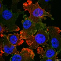
Anti-PD-L1, clone 5H1, Cat. No. MABC1115, is a mouse monoclonal antibody that detects PD-L1 and has been tested for use in Immunocytochemistry, Immunohistochemistry (Paraffin), and Western Blotting.
      Key Applications
      • Immunocytochemistry
      • Immunohistochemistry (Paraffin)
      • Western Blotting
      Application NotesImmunohistochemistry (Paraffin) Analysis: A representative lot detected PD-L1 in Immunohistochemistry applications (Bigelow, E., et. al. (2013). J Vis Exp. 3;(71); Spranger, S., et. al. (2013). Sci Transl Med. 5(200):200ra116; Parra, E.R., et. al. (2018). Appl Immunohistochem Mol Morphol. 26(2):83-93; Andorsky, D.J., et. al. (2011). Clin Cancer Res. 17(13):4232-44; Sunshine, J.C., et. al. (2017). Clin Cancer Res. 23(16):4938-4944).

      Western Blotting Analysis: A representative lot detected PD-L1 in Western Blotting applications (Parra, E.R., et. al. (2018). Appl Immunohistochem. Mol. Morphol. 26(2):83-93_.
      Biological Information
      ImmunogenFull-length human recombinant PD-L1.
      Epitopeextracellular domain
      Clone5H1
      ConcentrationPlease refer to lot specific datasheet.
      HostMouse
      SpecificityClone 5H1 detects human Programmed cell death ligand 1 (PD-L1). It targets an epitope with in the extracellular domain.
      IsotypeIgG1κ
      Species Reactivity
      • Human
      Species Reactivity NoteHuman.
      Antibody TypeMonoclonal Antibody
      Entrez Gene Number
      Gene Symbol
      • CD274
      • B7H1
      • PDCD1L1
      • PDCD1LG1
      • PDL1
      Purification MethodProtein G purified
      UniProt Number
      Molecular Weight33.28 kDa calculated.
      Physicochemical Information
      Dimensions
      Materials Information
      Toxicological Information
      Safety Information according to GHS
      Safety Information
      Product Usage Statements
      Quality AssuranceEvaluated by Immunocytochemistry in MDA-MB-231 cells.

      Immunocytochemistry Analysis: A 1:50 dilution of this antibody detected PD-L1 in MDA-MB-231 cells.
      Usage Statement
      • Unless otherwise stated in our catalog or other company documentation accompanying the product(s), our products are intended for research use only and are not to be used for any other purpose, which includes but is not limited to, unauthorized commercial uses, in vitro diagnostic uses, ex vivo or in vivo therapeutic uses or any type of consumption or application to humans or animals.
      Storage and Shipping Information
      Storage ConditionsStable for 1 year at -20°C from date of receipt. Handling Recommendations: Upon receipt and prior to removing the cap, centrifuge the vial and gently mix the solution. Aliquot into microcentrifuge tubes and store at -20°C. Avoid repeated freeze/thaw cycles, which may damage IgG and affect product performance.
      Packaging Information
      Material Size100 µL
      Transport Information
      Supplemental Information
      Specifications
      Global Trade Item Number
      카탈로그 번호 GTIN
      MABC1115-100UL 04054839613760

      관련 제품 및 어플리케이션