Millipore Sigma Vibrant Logo

MABF3111-25UG Anti-KIR3DL2/CD158k Antibody, clone Q66

MABF3111-25UG
25 μg  
가격 검색...
가격을 검색할 수 없습니다
Minimum Quantity needs to be mulitiple of
Maximum Quantity is
가격 문의 추가 정보
()이 할인됨
 
가격 문의
현재 재고 없음
현재 재고 없음
예상 출고 가능일 
단종품
제한된 수량 가능
재고여부 확인
    Remaining: will advise
      Remaining: will advise
      추천사항
      고객 서비스로 문의
      Contact Customer Service


      고객 서비스로 문의

      개요

      Replacement Information
      Description
      Catalogue NumberMABF3111-25UG
      DescriptionAnti-KIR3DL2/CD158k Antibody, clone Q66
      Alternate Names
      • Killer cell immunoglobulin-like receptor 3DL2
      • CD158 antigen-like family member K
      • Natural killer-associated transcript 4
      • NKAT-4
      • p70 natural killer cell receptor clone CL-5
      • p70 NK receptor CL-5
      • CD158kp70 NK receptor CL-5)
      Background InformationKiller cell immunoglobulin-like receptor 3DL2 (UniProt: P43630; also known as CD158 antigen-like family member K, Natural killer-associated transcript 4, NKAT-4, p70 natural killer cell receptor clone CL-5, p70 NK receptor CL-5, CD158k) is encoded by the KIR3DL2 (also known as CD158K, NKAT4) gene (Gene ID: 3812) in human. KIR3DL2 is a single-pass type I membrane glycoprotein, which is synthesized with a signal peptide (aa 1-21), which is subsequently cleaved off to produce the mature protein that contains an extracellular domain (aa 22-340), a transmembrane domain (aa 341-360), and a cytoplasmic domain (aa 361-455). It is expressed as a disulfide-linked homodimer and each chain contains three Ig-like domains (42-102; 137-202; and 237-300), and a long cytoplasmic tail that contains two ITIM motifs. Its expression is observed in astrocytes, NK cells, CD8+ T cells, and a small proportion of CD4+ T cells. Its expression is upregulated upon activation of NK and T cells. It serves as a receptor on NK cells and T cells for MHC class I molecules and upon binding of peptide-free HLA-F open conformer, it negatively regulates NK and T cell effector functions. Through interaction with HLA-F, it is suggested to protect motor neurons from astrocyte-induced toxicity. KIR3DL2 also binds to CpG-oligodeoxynucleotides (CpG-ODN), and this ligation induces KIR3DL2 down-modulation from the cell surface and translocation to the endosome to deliver the CpG-ODN to TLR9. Engagement of KIR3DL2 by ODN is reported to induce the internalization of the receptor and caspase-dependent apoptosis of malignant T cells. This apoptotic process is correlated to a dephosphorylation of STAT3 that is constitutively phosphorylated and activated in Se´zary cells. KIR3DL2 is considered as an important diagnostic and prognostic marker for the detection of the tumoral T cell burden of Se´zary syndrome patients. (Ref.: Schmitt, C., et al. (2017). Front. Immunol. 8; 1010; Ghazi, B., et al. (2015). J. Clin. Invest. 135(1); 229-237; Dorothee, G., et al. (2003). Oncogene. 22(46); 7192-7198; Pende, D., et al. (1996). J. Exp. Med. 184(2); 505-518).
      References
      Product Information
      FormatPurified
      PresentationPurified mouse monoclonal antibody IgM in PBS without azide.
      Quality LevelMQ200
      Applications
      ApplicationAnti-KIR3DL2/CD158k, clone Q66, Cat. No. MABF3111, is a mouse monoclonal antibody that detects KIR3DL2/CD158k and is tested for use in Flow Cytometry, Immunofluorescence, and Western Blotting.
      Key Applications
      • Flow Cytometry
      • Western Blotting
      • Immunofluorescence
      Application NotesTested Applications

      Western Blotting Analysis: A representative lot detected KIR3DL2/CD158k in Western Blotting application (Pende, D., et al. (1996). J Exp Med. 184(2):505-18).

      Flow Cytometry Analysis: A representative lot detected KIR3DL2/CD158k in Flow Cytometry applications (Pende, D., et al. (1996). J Exp Med. 184(2):505-18; Sivori, S., et al. (2002). Proc Natl Acad Sci USA. 99(7):4526-31; Dorothee, G., et al. (2003). Oncogene.;22(46):7192-8; Bahler, D.W., et al. (2008). Cytometry B Clin Cytom. 74(3):156-62; Carrega, P., et al. (20 9). PLoS One.;4(12):e8132; Falco, M., et al. (2010). J Immunol. 185(1):433-41; Ghazi, B., et al. (2015). J Invest Dermatol. 135(1):229-237).

      Immunofluorescence Analysis: A representative lot detected KIR3DL2/CD158k in Immunofluorescence applications (Ghazi, B., et al. (2015). J Invest Dermatol.;135(1):229-237).

      Note: Actual optimal working dilutions must be determined by end user as specimens, and experimental conditions may vary with the end user
      Biological Information
      ImmunogenNK cells clone DP7 (surface phenotype: CD3-CD16+CD56+GL183-EB6-Z27-).
      EpitopeExtracellular domain
      CloneQ66
      Concentration0.5 mg/mL. Please refer to guidance on suggested starting dilutions and/or titers per application and sample type.
      HostMouse
      SpecificityClone Q66 is a mouse monoclonal antibody that detects Killer cell immunoglobulin-like receptor 3DL2 (KIR3DL2). It targets an epitope within the extracellular domain.
      IsotypeIgMκ
      Species Reactivity
      • Human
      Species Reactivity NoteHuman.
      Antibody TypeMonoclonal Antibody
      Entrez Gene Number
      Gene Symbol
      • KIR3DL2
      Purification MethodLigatrap
      UniProt Number
      Molecular Weight50.23 kDa calculated.
      Physicochemical Information
      Dimensions
      Materials Information
      Toxicological Information
      Safety Information according to GHS
      Safety Information
      Product Usage Statements
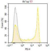
      Quality AssuranceEvaluated by Flow Cytometry in Human peripheral blood mononuclear cells (PBMC).

      Flow Cytometry Analysis: 1.0 µg of this antibody detected KIR3DL2/CD158k in one million Human peripheral blood mononuclear cells (PBMC).
      Usage Statement
      • Unless otherwise stated in our catalog or other company documentation accompanying the product(s), our products are intended for research use only and are not to be used for any other purpose, which includes but is not limited to, unauthorized commercial uses, in vitro diagnostic uses, ex vivo or in vivo therapeutic uses or any type of consumption or application to humans or animals.
      Storage and Shipping Information
      Storage ConditionsStore at -10°C to -25°C. Handling Recommendations: Upon receipt and prior to removing the cap, centrifuge the vial and gently mix the solution. Aliquot into microcentrifuge tubes and store at -20°C. Avoid repeated freeze/thaw cycles, which may damage IgG and affect product performance.
      Packaging Information
      Material Size25 μg
      Transport Information
      Supplemental Information
      Specifications
      Global Trade Item Number
      카탈로그 번호 GTIN
      MABF3111-25UG 04065270451774