Lipid mixtures containing a very high proportion of saturated fatty acids only modestly impair insulin signaling in cultured muscle cells.
Newsom, SA; Everett, AC; Park, S; Van Pelt, DW; Hinko, A; Horowitz, JF
PloS one
10
e0120871
2015
요약 표시
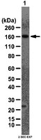
In vitro examinations of the effect of saturated fatty acids on skeletal muscle insulin action often use only one or two different fatty acid species, which does not resemble the human plasma fatty acid profile. We compared graded concentrations (0.1-0.8 mM) of 3 different lipid mixtures: 1) a physiologic fatty acid mixture (NORM; 40% saturated fatty acids), 2) a physiologic mixture high in saturated fatty acids (HSFA; 60% saturated fatty acids), and 3) 100% palmitate (PALM) on insulin signaling and fatty acid partitioning into triacylglycerol (TAG) and diacylglycerol (DAG) in cultured muscle cells. As expected, PALM readily impaired insulin-stimulated pAktThr308/Akt and markedly increased intracellular DAG content. In contrast, the fatty acid mixtures only modestly impaired insulin-stimulated pAktThr308M/Akt, and we found no differences between NORM and HSFA. Importantly, NORM and HSFA did not increase DAG content, but instead dose-dependently increased TAG accumulation. Therefore, the robust impairment in insulin signaling found with palmitate exposure was attenuated with physiologic mixtures of fatty acids, even with a very high proportion of saturated fatty acids. This may be explained in part by selective partitioning of fatty acids into neutral lipid (i.e., TAG) when muscle cells were exposed to physiologic lipid mixtures. | Western Blotting | 25793412
 |
Proteomic analysis of protein palmitoylation in adipocytes.
Ren, W; Jhala, US; Du, K
Adipocyte
2
17-28
2013
요약 표시
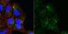
Protein palmitoylation, by modulating the dynamic interaction between protein and cellular membrane, is involved in a wide range of biological processes, including protein trafficking, sorting, sub-membrane partitioning, protein-protein interaction and cell signaling. To explore the role of protein palmitoylation in adipocytes, we have performed proteomic analysis of palmitoylated proteins in adipose tissue and 3T3-L1 adipocytes and identified more than 800 putative palmitoylated proteins. These include various transporters, enzymes required for lipid and glucose metabolism, regulators of protein trafficking and signaling molecules. Of note, key proteins involved in membrane translocation of the glucose-transporter Glut4 including IRAP, Munc18c, AS160 and Glut4, and signaling proteins in the JAK-STAT pathway including JAK1 and 2, STAT1, 3 and 5A and SHP2 in JAK-STAT, were palmitoylated in cultured adipocytes and primary adipose tissue. Further characterization showed that palmitoylation of Glut4 and IRAP was altered in obesity, and palmitoylation of JAK1 played a regulatory role in JAK1 intracellular localization. Overall, our studies provide evidence to suggest a novel and potentially regulatory role for protein palmitoylation in adipocyte function. | | 23599907
|
Sustained postexercise increases in AS160 Thr642 and Ser588 phosphorylation in skeletal muscle without sustained increases in kinase phosphorylation.
Schweitzer, GG; Arias, EB; Cartee, GD
Journal of applied physiology (Bethesda, Md. : 1985)
113
1852-61
2012
요약 표시
Prior exercise by rats can induce a sustained increase in muscle Akt substrate of 160 kDa (AS160) phosphorylation on Thr(642) (pAS160(Thr642)). Because phosphorylation of AS160 on both AS160(Thr642) and AS160(Ser588) is important for insulin-stimulated glucose transport (GT), we determined if exercise would also induce a sustained increase in pAS160(Ser588) concomitant with persistently elevated pAS160(Thr642) and GT. Given that the mechanisms for sustained postexercise (PEX) effects on pAS160 were uncertain, we also studied the four kinases known to phosphorylate AS160 (Akt, AMPK, RSK, and SGK1). In addition, because the serine/threonine phosphatase(s) that dephosphorylate muscle AS160 were previously unidentified, we assessed the ability of four serine/threonine phosphatases (PP1, PP2A, PP2B, and PP2C) to dephosphorylate AS160. We also evaluated exercise effects on posttranslational modifications (Tyr(307) and Leu(309)) that regulate PP2A. In isolated epitrochlearis muscles from rats, GT at 3hPEX with insulin significantly (P less than 0.05) exceeded SED controls. Muscles from 0hPEX vs. 0hSED and 3hPEX vs. 3hSED rats had greater pAS160(Thr642) and pAS160(Ser588). AMPK was the only kinase with greater phosphorylation at 0hPEX vs. 0hSED, and none had greater phosphorylation at 3hPEX vs. 3hSED. Each phosphatase was able to dephosphorylate pAS160(Thr642) and pAS160(Ser588) in cell-free assays. Exercise did not alter posttranslational modifications of PP2A. Our results revealed: 1) pAMPK as a potential trigger for increased pAS160(Thr642) and pAS160(Ser588) at 0hPEX; 2) PP1, PP2A, PP2B, and PP2C were each able to dephosphorylate AS160; and 3) sustained PEX-induced elevations of pAS160(Thr642) and pAS160(Ser588) were attributable to mechanisms other than persistent phosphorylation of known AS160 kinases or altered posttranslational modifications of PP2A. | | 22936728
|
Activity profiles of cholinergic and intermingled GABAergic and putative glutamatergic neurons in the pontomesencephalic tegmentum of urethane-anesthetized rats.
Soufiane Boucetta,Barbara E Jones
The Journal of neuroscience : the official journal of the Society for Neuroscience
29
2009
요약 표시
Cholinergic neurons in the pontomesencephalic tegmentum form part of the ascending activating system and are thought to participate in stimulating cortical activation. Yet in the laterodorsal tegmental and pedunculopontine tegmental nuclei (LDT and PPT), they lie intermingled with GABAergic and glutamatergic neurons, which could also modulate cortical activity and sleep-wake state. To characterize the discharge of these cell types in relation to cortical activity, we recorded neurons in urethane-anesthetized rats during spontaneous slow wave and somatosensory evoked fast electroencephalographic (EEG) activity, then labeled the cells by juxtacellular technique with Neurobiotin (Nb) and dual-immunostained them for vesicular acetylcholine transporter (VAChT) and glutamic acid decarboxylase (GAD). All cholinergic cells discharged minimally during prestimulation (approximately 0.5 Hz) and moderately in a tonic manner (approximately 4 Hz) during stimulation. Being heterogeneous, some GABAergic, called On, cells (approximately 48%) increased their discharge (from approximately 4 to 7 Hz), whereas others, called Off cells (approximately 38%), decreased or ceased firing during stimulation. Similarly, some noncholinergic/non-GABAergic On cells increased (from approximately 2 to 6 Hz, approximately 49%), whereas other Off cells decreased firing ( approximately 35%) during stimulation. Putative glutamatergic On together with GABAergic On neurons could thus act in parallel with cholinergic cells to stimulate cortical activation. Possibly influenced by cholinergic On and glutamatergic Off cells, whose change in discharge precedes theirs, the GABAergic Off cells could oppose neighboring neurons such as noradrenergic cells, which discharge during waking and cease firing during sleep. By concerted activity, these heterogeneous cell groups can modulate cortical activity and behavioral state across the sleep-waking cycle. | | 19357291
|