Custom Premix Selecting "Custom Premix" option means that all of the beads you have chosen will be premixed in manufacturing before the kit is sent to you.
Catalogue Number
Ordering Description
Qty/Pack
List
この品目はお気に入りに追加されました。
動物種
パネルタイプ
選択したキット
数量
カタログ番号
注文内容
Qty/Pk
価格
96-Well Plate
数量
カタログ番号
注文内容
Qty/Pk
価格
その他の試薬を追加 (MAPmatesとともに使用するにはバッファーおよび検出キットが必要です)
数量
カタログ番号
注文内容
Qty/Pk
価格
48-602MAG
Buffer Detection Kit for Magnetic Beads
1 Kit
Space Saver Option(チェックを入れると箱詰めから袋詰めに変更となりますのでご注意ください) Customers purchasing multiple kits may choose to save storage space by eliminating the kit packaging and receiving their multiplex assay components in plastic bags for more compact storage.
Recognizes the ~55 kDa c-Fos protein phosphorylated at Ser374. Does not detect the unphosphorylated form.
Catalogue Number
ST1029
Brand Family
Calbiochem®
Application Data
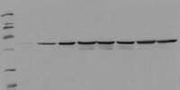
Detection of human Fos, phosphorylated on Ser374, by immunoblotting. Samples: Serum starved HepG2 cells incubated with EGF for 0 min (lane 1), 5 min (lane 2), 15 min (lane 3), 30 min (lane 4), 1 h (lane 5), 2 h (lane 6), 4 h (lane 7), and 8 h (lane 8). Primary antibody: PhosphoDetect™ Anti-Fos (pSer374) Mouse mAb (34E4) (Cat. No. ST1029) (0.5 µg/ml). Detection: chemiluminescence.
References
References
Chen, R-H., et al. 1993. Proc. Natl. Acad. Sci. USA90, 10952.
Product Information
Form
Lyophilized
Formulation
100 µg antibody lyophilized from 2X PBS, PEG, sucrose and 200 µl lyophilized control lysate from EGF-treated HEPG2 cells.
Does not detect the unphosphorylated form of c-fos. For immunoblotting use 20 µl control lysate per lane (minigel) for chemiluminescent detection. Antibody should be titrated for optimal results in individual systems.
Biological Information
Immunogen
a synthetic phosphopeptide corresponding to amino acids surrounding the Ser³⁷⁴ phosphorylation site of human c-Fos
Immunogen
Human
Clone
34E4
Host
Mouse
Isotype
IgG₁
Species Reactivity
Human
Antibody Type
Monoclonal Antibody
Physicochemical Information
Dimensions
Materials Information
Toxicological Information
Safety Information according to GHS
Safety Information
Product Usage Statements
Storage and Shipping Information
Ship Code
Shipped with Blue Ice or with Dry Ice
Toxicity
Irritant
Storage
-20°C
Avoid freeze/thaw
Avoid freeze/thaw
Do not freeze
Ok to freeze
Special Instructions
Reconstitute antibody with 1 ml water (15 minutes, room temperature). Following reconstitution, aliquot and freeze in liquid nitrogen. Reconstituted antibody can be stored at -80°C for up to 1 year. Aliquots may be stored at 4°C for up to 3 months and should be thawed at 37°C. Reconstitute control lysate with 200 µl H₂O. After complete solubilization of the proteins add 200 µl SDS-PAGE sample buffer and incubate at 90°C for 5 min. Following reconsitution aliquot and freeze (-20°C). Avoid freeze/thaw cycles.
Packaging Information
Transport Information
Supplemental Information
Specifications
Global Trade Item Number
カタログ番号
GTIN
ST1029-1SETCN
04055977208689
Documentation
PhosphoDetect™ Anti-Fos (pSer³⁷⁴) Mouse mAb (34E4) (M)SDS
PhosphoDetect™ Anti-Fos (pSer³⁷⁴) Mouse mAb (34E4) 試験成績書(CoA)
タイトル
ロット番号
ST1029
参考資料
参考資料の概要
Chen, R-H., et al. 1993. Proc. Natl. Acad. Sci. USA90, 10952.
データシート
Note that this data sheet is not lot-specific and is representative of the current specifications for this product. Please consult the vial label and the certificate of analysis for information on specific lots. Also note that shipping conditions may differ from storage conditions.
Detection of human Fos, phosphorylated on Ser374, by immunoblotting. Samples: Serum starved HepG2 cells incubated with EGF for 0 min (lane 1), 5 min (lane 2), 15 min (lane 3), 30 min (lane 4), 1 h (lane 5), 2 h (lane 6), 4 h (lane 7), and 8 h (lane 8). Primary antibody: PhosphoDetect™ Anti-Fos (pSer374) Mouse mAb (34E4) (Cat. No. ST1029) (0.5 µg/ml). Detection: chemiluminescence.
Description
Mouse monoclonal antibody purified from serum-free cell culture supernatant by thiophilic adsorption and size exclusion chromatography. Supplied with a positive control lysate consisting of EGF-treated HepG2 cells. Recognizes the ~55 kDa c-fos protein phosphorylated at Ser374 by MAP kinase.
Background
The early gene product c-Fos is expressed following mitogenic stimulation and functions as a sensor for MAPK signal duration. When MAPK activation is transient, MAPK activity declines before accumulation of the c-Fos protein. When MAPK activation is sustained, c-Fos is phosphorylated by MAPK at Ser374. Phosphorylation stabilizes the Fos protein and primes c-Fos for additional phosphorylation at Thr325.
Host
Mouse
Immunogen species
Human
Immunogen
a synthetic phosphopeptide corresponding to amino acids surrounding the Ser³⁷⁴ phosphorylation site of human c-Fos
Clone
34E4
Isotype
IgG₁
Species
human
Positive control
HepG2 cells treated with EGF
Form
Lyophilized
Formulation
100 µg antibody lyophilized from 2X PBS, PEG, sucrose and 200 µl lyophilized control lysate from EGF-treated HEPG2 cells.
Preservative
≤0.1% sodium azide (antibody only)
Comments
Does not detect the unphosphorylated form of c-fos. For immunoblotting use 20 µl control lysate per lane (minigel) for chemiluminescent detection. Antibody should be titrated for optimal results in individual systems.
Storage
Avoid freeze/thaw
-20°C
Do Not Freeze
Ok to freeze
Special Instructions
Reconstitute antibody with 1 ml water (15 minutes, room temperature). Following reconstitution, aliquot and freeze in liquid nitrogen. Reconstituted antibody can be stored at -80°C for up to 1 year. Aliquots may be stored at 4°C for up to 3 months and should be thawed at 37°C. Reconstitute control lysate with 200 µl H₂O. After complete solubilization of the proteins add 200 µl SDS-PAGE sample buffer and incubate at 90°C for 5 min. Following reconsitution aliquot and freeze (-20°C). Avoid freeze/thaw cycles.
Toxicity
Irritant
References
Chen, R-H., et al. 1993. Proc. Natl. Acad. Sci. USA90, 10952.