
ECM590
Blood Vessel Staining Kit
Reagents & materials supplied in the Blood Vessel Staining Kit are sufficient for staining 100 tissue sections & when stored at 2-8ºC is stable up to the expiration date.
製品名
Blood Vessel Staining Kit, Reagents & materials supplied in the Blood Vessel Staining Kit are sufficient for staining 100 tissue sections & when stored at 2-8ºC is stable up to the expiration date.
species reactivity
human
manufacturer/tradename
Chemicon®
technique(s)
immunohistochemistry (formalin-fixed, paraffin-embedded sections): suitable
detection method
colorimetric
shipped in
wet ice
Quality Level
Application
Cell Structure
Disclaimer
General description
Background:
Angiogenesis and tumor-associated neovascularization are complex processes by which new blood vessels are formed from pre-existing vasculature. Several studies implicate tumor-associated blood vessel formation as a central part in the process of growth, invasion and metastasis of malignancies (Rak et al., 1995; Jahroudi & Greenberger, 1995; Saphir, 1997).
The most widely used endothelial cell markers for studying angiogenesis/neovascularization are von Willebrand Factor (Factor VIII Related Antigen) and CD31 (PECAM-1). von Willebrand Factor is synthesized by endothelial cells, causing adhesion of platelets to injured vessel walls and functions as a carrier and stabilizer for coagulation of Factor VIII. CHEMICON′s von Willebrand Factor antibody reacts specifically with the endothelial cells of blood vessels and is a useful marker for the identification of endothelial lineage in tumors. It is important to note that not all endothelial cells synthesize von Willebrand factor, therefore a small percentage of these vascular-rich tumors might not stain with this antibody. CD31 or platelet endothelial adhesion molecule (PECAM-1), is a type I integral membrane glycoprotein and a member of the immunoglobulin super family of cell surface receptors. It is strongly expressed by all endothelial cells and weakly by several types of leucocytes (Parums et al., 1990). According to several studies, staining of blood vessels with CD31 antibodies has shown to be suitable for the identification of angiogenesis in several types of malignancies such as lung (Giatromanolaki et al., 1996), breast (Horak et al., 1992) and colorectal cancer (Takebayashi et al., 1996). CHEMICON′s monoclonal antibody to CD31 is very specific for the staining of vascular endothelial cells in normal and cancerous tissue.
CHEMICON′s Blood Vessel Staining Kit is a very sensitive immunohistochemical tool for the detection of vascular endothelial cells, containing both anti-von Willebrand Factor and anti-CD31 primary antibodies and is applicable to a variety of species. This immunoperoxidase kit provides a simple, all-in-one method for the staining of frozen and paraffin-embedded sections in human (both CD31 and VWF) or rat and mouse (VWF only) tissues
Other Notes
Mouse anti-CD31 Monoclonal Antibody (Part No. 90214): One vial containing 100 μL at 15 mg/mL (total protein including IgG and Carrier protein).
Goat anti-Rabbit Secondary Antibody (Part No. 21537): One 15 mL ready-to-use solution containing biotinylated goat anti-rabbit IgG in PBS with a carrier protein.
Goat anti-Mouse Secondary Antibody (Part No. 21538): One 15 mL ready-to-use solution containing biotinylated goat anti-mouse IgG in PBS with a carrier protein.
Blocking Reagent (Part No. 20783): One 15 mL ready-to-use solution containing normal goat serum in PBS with a carrier protein.
Streptavidin-HRP (Part No. 90215): One 15 ml ready-to-use solution of Streptavidin-HRP diluted in Tris-buffered saline (TBS).
DAB Chromogen A (Part No. 90216): One 5 mL bottle of 3, 3′ Diaminobenzidine diluted in TBS.
DAB Chromogen B (Part No: 90217): Two 12 mL bottles of hydrogen peroxide diluted in distilled water.
20X Rinse Buffer (Part No. 90218): One 275 mL bottle of 20X TBS.
Packaging
Preparation Note
Legal Information
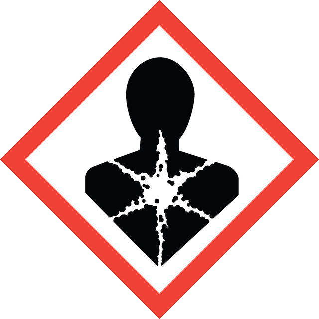

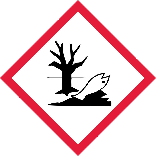
signalword
Danger
Hazard Classifications
Aquatic Chronic 2 - Carc. 1B - Eye Irrit. 2 - Muta. 2 - Skin Irrit. 2 - Skin Sens. 1
保管分類
6.1C - Combustible acute toxic Cat.3 / toxic compounds or compounds which causing chronic effects
wgk
WGK 3
適用法令
試験研究用途を考慮した関連法令を主に挙げております。化学物質以外については、一部の情報のみ提供しています。 製品を安全かつ合法的に使用することは、使用者の義務です。最新情報により修正される場合があります。WEBの反映には時間を要することがあるため、適宜SDSをご参照ください。
Please refer to KIT Component information
pdsc
Please refer to KIT Component information
prtr
Please refer to KIT Component information
fsl
Please refer to KIT Component information
ishl_indicated
Please refer to KIT Component information
ishl_notified
Please refer to KIT Component information
cart
キットコンポーネントの情報を参照してください
jan
試験成績書(COA)
製品のロット番号・バッチ番号を入力して、試験成績書(COA) を検索できます。ロット番号・バッチ番号は、製品ラベルに「Lot」または「Batch」に続いて記載されています。
資料
Cell based angiogenesis assays to analyze new blood vessel formation for applications of cancer research, tissue regeneration and vascular biology.
関連コンテンツ
"The successful, reliable culture of epithelial cells is critical for many areas of research, including dermatology, respiratory research, and cancer research. Because the breakdown of control mechanisms in epithelial cells is a frequent contributor to cancer progression and metastasis, epithelial cell culture is particularly important for cancer research. EpiGRO™ media formulations are optimized to provide better viability, proliferation rates, morphology and culture stability than other commercially available options. The media are provided in unique, light-blocking, temperature-monitored packaging to ensure stability and protect the media from damage by light, contamination, and excessive heat. The media do not require or contain any antimicrobials or phenol red. These components can cause cell stress and influence experimental results by masking the true performance or health of the cell culture. Phenol red acts like an estrogen and may stimulate growth independently of experimental variables. "
グローバルトレードアイテム番号
| カタログ番号 | GTIN |
|---|---|
| ECM590 | 04053252287077 |
ライフサイエンス、有機合成、材料科学、クロマトグラフィー、分析など、あらゆる分野の研究に経験のあるメンバーがおります。.
製品に関するお問い合わせはこちら(テクニカルサービス)