Millipore Sigma Vibrant Logo

ABN1649 Anti-TMC1

View This Product on Sigma-Aldrich
ABN1649
100 µL  
価格を検索中…
価格が見つかりません
Minimum Quantity needs to be mulitiple of
Maximum Quantity is
弊社照会 詳細を表示 
値引
()
 
弊社照会
現在国内在庫なし
現在国内在庫なし
現在国内在庫有り 
販売中止
在庫僅少
現在国内在庫あり
    Remaining : Will advise
      Remaining : Will advise
      注文対象外
      お問合せください
      Contact Customer Service


      お問合せください

      概要

      Replacement Information

      主要スペック表

      Species ReactivityKey ApplicationsHostFormatAntibody Type
      M, MkIF, ICC, WBRbSerumPolyclonal Antibody
      Description
      Catalogue NumberABN1649
      DescriptionAnti-TMC1
      Alternate Names
      • Transmembrane channel-like protein 1
      • Beethoven protein
      • Deafness protein
      • Transmembrane cochlear-expressed protein 1
      Background InformationTransmembrane channel-like protein 1 (UniProt: Q8R4P5; also known as Beethoven protein, Deafness protein, Transmembrane cochlear-expressed protein 1, TMC1) is encoded by the Tmc1 (also known as Bth, dn) gene (Gene ID: 13409) in murine species. TMC1 is a multi-pass membrane protein that forms ion channel required for the normal function of cochlear hair cells. It is detected in cochlear inner and outer hair cells and in neurosensory epithelia of the vestibular end organs. Its expression is also reported in cortex, cerebellum, eye, colon, ovary, and testis. It is expressed at low levels in temporal bone from embryonic day 14 to day 1 after birth in mice and increases significantly (8 to 16 fold) at day 5, 10 and 20 and then continues to be expressed up to day 90. Two isoforms of TMC1 have been reported in mice that are produced by alternative splicing. Mutations in Tmc1 gene can cause hearing loss in humans and mice. Heterozygotes show progressive hair-cell degeneration from day 20 onwards in mice, leading to severe depletion of inner hair cells and scattered loss of outer hair cells, and progressive loss of the Preyer reflex from around day 30. Homozygotes show almost complete degeneration of inner hair cells, and little or no Preyer reflex at any age.
      References
      Product Information
      FormatSerum
      PresentationRabbit polyclonal antiserum with 0.05% sodium azide.
      Quality LevelMQ100
      Applications
      ApplicationAnti-TMC1, Cat. No. ABN1649, is a highly specific rabbit polyclonal antibody that targets Transmembrane channel-like protein 1 and has been tested for use in Immunocytochemistry, Immunofluorescence, and Western Blotting.
      Key Applications
      • Immunofluorescence
      • Immunocytochemistry
      • Western Blotting
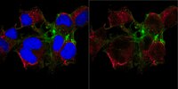
      Application NotesImmunocytochemistry Analysis: A 1:100 dilution from a representative lot detected TMC1 in COS-7 cells.

      Immunofluorescence Analysis: A representative lot detected TMC1 in P9 mouse outer hair cells (OHCs) (Kurima, K., et. al. (2015). Cell Rep. 12(10):1606-17).

      Western Blotting Analysis: A representative lot detected TMC1 in COS-7 cells (Labay, V., et. al. (2010). Biochemistry. 49(39):8592-8).

      Western Blotting Analysis: A 1:1,000 dilution from a representative lot detected TMC1 in HEK293 cells transiently transfected with Fugene and TMCx-cherry cDNAs (Courtesy of Dr. Angela Ballesteros).

      Immunofluorescence Analysis: A representative lot detected TMC1 in COS-7 transfected cells expressing TMC1-GFP (Courtesy of Dr. Angela Ballesteros).
      Biological Information
      ImmunogenKLH-conjugated linear peptide corresponding to 20 amino acids from the N-terminal cytoplasmic domain of murine Transmembrane channel-like protein 1.
      Epitopecytoplasmic domain
      ConcentrationPlease refer to lot specific datasheet.
      HostRabbit
      SpecificityThis rabbit polyclonal antibody detects Transmembrane channel-like protein 1 in murine retina. It targets an epitope within 20 amino acids from the N-terminal region.
      IsotypeIgG
      Species Reactivity
      • Mouse
      • Monkey
      Species Reactivity NoteMonkey, Mouse.
      Antibody TypePolyclonal Antibody
      Entrez Gene Number
      Gene Symbol
      • Tmc1
      • Bth
      • dn
      Purification MethodUnpurified
      UniProt Number
      Molecular Weight~90 kDa observed; 87.26 kDa calculated. Uncharacterized bands may be observed in some lysate(s).
      Physicochemical Information
      Dimensions
      Materials Information
      Toxicological Information
      Safety Information according to GHS
      Safety Information
      Product Usage Statements
      Quality AssuranceEvaluated by Western Blotting in mouse retinal tissue lysate.

      Western Blotting Analysis: A 1:500 dilution of this antibody detected TMC1 in 10 µg of mouse retinal tissue lysate.
      Usage Statement
      • Unless otherwise stated in our catalog or other company documentation accompanying the product(s), our products are intended for research use only and are not to be used for any other purpose, which includes but is not limited to, unauthorized commercial uses, in vitro diagnostic uses, ex vivo or in vivo therapeutic uses or any type of consumption or application to humans or animals.
      Storage and Shipping Information
      Storage ConditionsStable for 1 year at -20°C from date of receipt. Handling Recommendations: Upon receipt and prior to removing the cap, centrifuge the vial and gently mix the solution. Aliquot into microcentrifuge tubes and store at -20°C. Avoid repeated freeze/thaw cycles, which may damage IgG and affect product performance.
      Packaging Information
      Material Size100 µL
      Transport Information
      Supplemental Information
      Specifications
      Global Trade Item Number
      カタログ番号 GTIN
      ABN1649 04054839355844