Millipore Sigma Vibrant Logo

04-1481-25UG Anti-RPA2 p34 Antibody, clone RPA20, 1-46 (mouse monoclonal)

View This Product on Sigma-Aldrich
04-1481-25UG
25 μg  
価格を検索中…
価格が見つかりません
Minimum Quantity needs to be mulitiple of
Maximum Quantity is
弊社照会 詳細を表示 
値引
()
 
弊社照会
現在国内在庫なし
現在国内在庫なし
現在国内在庫有り 
販売中止
在庫僅少
現在国内在庫あり
    Remaining : Will advise
      Remaining : Will advise
      注文対象外
      お問合せください
      Contact Customer Service

      Special Offers[海外情報]


      お問合せください

      概要

      Replacement Information

      Special Offers[海外情報]

      主要スペック表

      Species ReactivityKey ApplicationsHostFormatAntibody Type
      H, MWBMPurifiedMonoclonal Antibody
      Description
      Catalogue Number04-1481-25UG
      DescriptionAnti-RPA2 p34 Antibody, clone RPA20, 1-46 (mouse monoclonal)
      Alternate Names
      • RF-A protein 2
      • RP-A p32
      • RP-A p34
      • Replication factor A protein 2
      • replication protein A2 (32kD)
      • replication protein A2, 32kDa
      • Replication protein A 32kDa subunit
      • RF-A, replication factor-A protein 2
      • p32
      • p34
      • RPA2
      • REPA2
      • RPA32.
      Background InformationRPA is a heterotrimeric protein complex that binds specifically to single-stranded DNA (ssDNA). It is composed of three subunits, RPA1 (70 kDa), RPA2 (32 kDa), and RPA3 (14 kDa), and plays multiple roles in DNA metabolism. RPA is required for DNA replication initiation, as well as replication elongation. At the onset of DNA replication, RPA is loaded onto chromatin after the binding of Cdc45 to origins. RPA is needed for subsequent loading of DNA polymerase and other replication proteins to initiate DNA replication. After DNA replication begins, RPA moves with replication forks, stabilizing ssDNA and assisting in DNA synthesis. In addition to its replication function, RPA is also known to play essential roles in damage repair and recombination.The 32 kDa subunit, is phosphorylated by the cdc2 family of kinases when cells enter S-phase and in response to DNA damage by ATM, ATR, and DNA-PK. Alternate names for RPA32 include replication protein A 32 kDa subunit, RP-A, RF-A, replication factor-A protein 2, p32, p34, RPA2, REPA2, and RPA32.
      References
      Product Information
      FormatPurified
      Control
      • HeLa cell lysate
      PresentationPurified mouse monoclonal IgG1 in buffer containing 0.1 M Tris-Glycine (pH 7.4, 150 mM NaCl) with 0.05% sodium azide.
      Quality LevelMQ100
      Applications
      ApplicationAnti-RPA2 p34 Antibody, clone RPA20 1-46 is a Mouse Monoclonal Antibody for detection of RPA2 p34 also known as Replication factor A protein 2 & has been validated in WB.
      Key Applications
      • Western Blotting
      Application NotesWestern Blot (SNAP ID) Analysis: 0.5 µg/mL from a previous lot detected RPA2 on 10 µg of NIH/3T3 cell lysate.
      Biological Information
      ImmunogenHis-tagged recombinant protein corresponding to human RPA2.
      EpitopeUnknown
      CloneRPA20, 1-46
      ConcentrationPlease refer to lot specific datasheet.
      HostMouse
      SpecificityThis antibody recognizes RPA2.
      IsotypeIgG1κ
      Species Reactivity
      • Human
      • Mouse
      Species Reactivity NoteProven to react with human and mouse.
      Antibody TypeMonoclonal Antibody
      Entrez Gene Number
      Gene Symbol
      • RPA2
      • REPA2
      • RPA32
      • RPA34
      Purification MethodProtein G Purified
      UniProt Number
      UniProt SummaryFUNCTION: Required for DNA recombination, repair and replication. The activity of RP-A is mediated by single-stranded DNA binding and protein interactions.

      SUBUNIT STRUCTURE: Heterotrimer of 70, 32 and 14 kDa chains. The DNA-binding activity may reside exclusively on the 70 kDa subunit. Binds to SERTAD3/RBT1. Interacts with TIPIN.

      SUBCELLULAR LOCATION: Nucleus. Note: Also present in PML nuclear bodies. Redistributes to discrete nuclear foci upon DNA damage.

      PTM: Phosphorylated in a cell-cycle-dependent manner (from the S phase until mitosis). Phosphorylated by ATR upon DNA damage, which promotes its translocation to nuclear foci. Can be phosphorylated in vitro by PRKDC/DNA-PK in the presence of Ku and DNA, and by CDC2.
      Molecular Weight~32 kDa
      Physicochemical Information
      Dimensions
      Materials Information
      Toxicological Information
      Safety Information according to GHS
      Safety Information
      Product Usage Statements
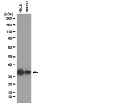
      Quality AssuranceEvaluated by Western Blot in HeLa cell lysate.

      Western Blot Analysis: 0.5 µg/mL of this antibody detected RPA2 on 10 µg of HeLa cell lysate.
      Usage Statement
      • Unless otherwise stated in our catalog or other company documentation accompanying the product(s), our products are intended for research use only and are not to be used for any other purpose, which includes but is not limited to, unauthorized commercial uses, in vitro diagnostic uses, ex vivo or in vivo therapeutic uses or any type of consumption or application to humans or animals.
      Storage and Shipping Information
      Storage ConditionsStable for 1 year at 2-8°C from date of receipt.
      Packaging Information
      Material Size25 μg
      Transport Information
      Supplemental Information
      Specifications
      Global Trade Item Number
      カタログ番号 GTIN
      04-1481-25UG 04054839350108