


ST1517 Sigma-AldrichAnti-PIP5K3 Mouse mAb (6C7)
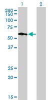

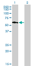
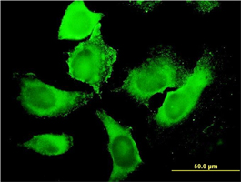
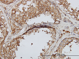
This Anti-PIP5K3 Mouse mAb (6C7) is validated for use in ELISA, Immunoblotting, Immunocytochemistry, Paraffin Sections for the detection of PIP5K3.
More>> This Anti-PIP5K3 Mouse mAb (6C7) is validated for use in ELISA, Immunoblotting, Immunocytochemistry, Paraffin Sections for the detection of PIP5K3. Less<<Anti-PIP5K3 Mouse mAb (6C7): のSDS(安全データシート)、CoA(試験成績書)およびCoQ(品質証明書)、カタログなど製品関連の技術資料を入手いただけます。
同義語: Anti-Phosphatidylinosi-3-Phosphate-Phosphatidylinosi-5-Kinase Type 3
お勧めの製品


概要
| Replacement Information |
|---|
主要スペック表
| Species Reactivity | Host | Antibody Type |
|---|---|---|
| H | M | Monoclonal Antibody |
価格&在庫状況
| カタログ番号 | 在庫状況 | 包装 | Qty/Pk | 価格 | 数量 | |
|---|---|---|---|---|---|---|
| ST1517-100UGCN |
|
100 μg |
|
— |
| References | |
|---|---|
| References | Tsuruta, F., et al. 2009. J. Cell Biol. 187 279. |
| Product Information | |
|---|---|
| Form | liquid |
| Formulation | In PBS, pH 7.2. |
| Negative control | 293T cells |
| Positive control | Human testes tissue, HeLa cells |
| Preservative | None |
| Quality Level | MQ100 |
| Physicochemical Information |
|---|
| Dimensions |
|---|
| Materials Information |
|---|
| Toxicological Information |
|---|
| Safety Information according to GHS |
|---|
| Safety Information |
|---|
| Product Usage Statements |
|---|
| Packaging Information |
|---|
| Transport Information |
|---|
| Supplemental Information |
|---|
| Specifications |
|---|
| Global Trade Item Number | |
|---|---|
| カタログ番号 | GTIN |
| ST1517-100UGCN | 04055977224184 |
Documentation
Anti-PIP5K3 Mouse mAb (6C7) (M)SDS
| タイトル |
|---|
参考資料
| 参考資料の概要 |
|---|
| Tsuruta, F., et al. 2009. J. Cell Biol. 187 279. |