Nitric oxide (NO) expression during annual reproductive activity in buffalo epididymis: a histochemical and immunocytochemical study.
Gaetano Scala,Lucianna Maruccio
Theriogenology
78
2012
概要を表示する
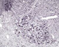
The buffalo is one of the few domestic animals that has a seasonal mating cycle, influenced by the photoperiod. It is known that the photoperiod regulates gonadal function probably via the pineal and/or hypothalamus-pituitary axis. Moreover, the hypothalamus (melatonin) and gonads influence the production of the signaling transmitter nitric oxide (NO), suggesting that the NO may have an important role in the regulation of gonadotropin-releasing hormone secretion. This further suggests the hypothesis that NO in the epididymis has an important role in the maturation of spermatozoa and their motility and posterior fertilization capacity. The aim of the present study is to investigate the seasonal variations in the morphology of the epididymis by means histochemical and immunocytochemical techniques. We used the NADPH-d, nitric oxide synthase (NOS) I and NOS III to clarify the relationship between epididymis function and NO signaling activity. The results of this work show that NO is present in the caput of epididymis during short photoperiods, i.e., periods of maximum gonadal activity (winter) and absent during long photoperiods, i.e., periods of gonadal regression according to the previously described role of NO in spermatozoa capacitation and motility in the caput epididymis. | 22406309
 |
Molecular typing of epithelial ovarian carcinomas using inflammatory markers.
Rouba Ali-Fehmi,Assaad Semaan,Sima Sethi,Haitham Arabi,Sudeshna Bandyopadhyay,Yaser R Hussein,Michael P Diamond,Ghasan Saed,Robert T Morris,Adnan R Munkarah
Cancer
117
2011
概要を表示する
Ovarian epithelial carcinomas have recently been classified as slow growing type I tumors and rapidly growing highly aggressive type II tumors. The present study sought to molecularly characterize type I and II tumors using known molecular markers. | 20818651
 |
Structural, histochemical and immunocytochemical study of the forestomach mucosa in domestic ruminants.
Scala G, Corona M, Maruccio L
Anatomia, histologia, embryologia
40
47-54. doi
2011
概要を表示する
The forestomach plays an important role in the digestion physiology of ruminants. The aim of this study is to clarify the morpho-functional role of the mucosa in each of the three compartments of the forestomach in three domestic ruminants species, viz cattle, buffalo and sheep, by means of structural, histochemical and immunocytochemical methods, including transmission electron microscopy, light microscopy and scanning electron microscopy. These methods were chosen to demonstrate the indirect evidence for the presence of nitric oxide (NO) employing NADPHd and nitric oxide synthase I (NOS I). The various cell layers of the forestomach epithelium are described and illustrated in detail. An intense NADPHd staining was observed in the granulosa, spinosa and basal layers of the epithelium, in particular in the cytoplasm over the nucleus. NOS I immunoreactivity was found in all specimens of the forestomach mucosa. The results of this study might reflect a possible role of NO in delaying the onset of cellular apoptosis in the forestomach mucosa of the domestic ruminants, by playing a role in the production of cell energy. | 21029150
 |
Microvasculature of the buffalo (Bubalus bubalis) choroid plexuses: structural, histochemical, and immunocytochemical study.
Scala G, Corona M, Langella E, Maruccio L
Microsc Res Tech
74
67-75.
2011
概要を表示する
The choroid plexuses (CPs) in mammals produce the cerebrospinal fluid (CSF). In the literature, the morphology of CPs and the process that regulates the production of CSF are virtually nonexistent for domestic ruminants. Thus this study has two aims: 1. to investigate the morpho-structure of the buffalo CP microvasculature utilizing light microscopy (LM), scanning electron microscopy (SEM) and transmission electron microscopy (TEM) techniques, and 2. to investigate the relationship between the blood vessels and both the elongated cells and the cells with multiple protrusions located in the CPs. SEM and TEM analyses of the CPs from buffalo brain showed morphological and structural features similar those reported in other mammalian species. Moreover the blood microvasculature is the major component responsible for the formation of the CSF, secreted by the encephalic CPs. In addition the chemical composition of this fluid depends on several morpho-functional characteristics of the vascularization of the CPs. These characteristics are as follows: two shapes of the vascular organization: lamina-like and ovoid-like elongated cells of the CPs, which connect the ventricular cavities to the blood capillaries; and the CP capillaries have diverse forms. In the present study the employment of NADPHd and NOS I was taken as indirect evidence for the presence of NO for investigation their specific role in CPs. Then NOS I immunoreactivity is found in the walls of CP blood vessels demonstrating indirectly the presence of NO with a vaso-dilatatory and autoregulation function of vascular tone by cholinergic nerve stimulation of blood vessel smooth muscle.© 2010 Wiley-Liss, Inc. | 21181712
 |
Lateralization of hippocampal nitric oxide mediator system in people with Alzheimer disease, multi-infarct dementia and schizophrenia.
Zdena Kristofiková,Iryna Kozmiková,Petra Hovorková,Jan Rícný,Petr Zach,Emmerich Majer,Jan Klaschka,Daniela Rípová
Neurochemistry international
53
2008
概要を表示する
There is evidence that brain lateralization underlying hemispheric specialization can be observed also at biochemical level. However, hemispheric differences in nitric oxide mediator system have not yet been evaluated. The hippocampus and planum temporale are highly asymmetrical regions but the degree of their laterality is altered in demented or psychotic people. In the study, l-glutamate/l-arginine/l-citrulline concentrations, nitric oxide synthase activities/expressions and nitrites/nitrates levels were estimated in autoptic hippocampi. Right/left laterality in endothelial synthase activity and in nitrites/nitrates was observed in controls. Lateral changes were estimated in patients with Alzheimer disease (a marked increase in activities of constitutive synthases and in expression of inducible enzyme in the left side) and schizophrenia (an increase in activities of all enzymes especially in the right side). Significant shifts from positive to negative correlations were found between laterality of some components of nitric oxide pathway and of planum temporale volumetry under pathological conditions. The hippocampal nitric oxide system appears to be globally right/left lateralized, especially via actions of highly asymmetrical endothelial synthase. The results suggest a specific involvement of all synthases in the development of selected diseases and show that lateral analyses are of sufficient sensitivity to reveal subtle links. The volumetric asymmetry of the planum temporale as a marker of handedness is not probably simply linked to brain laterality at biochemical level but reflects alterations due to pathological processes. | 18647632
 |
Distribution of neuronal nitric oxide synthase-immunoreactive neurons in the cerebral cortex and hippocampus during postnatal development.
Yoon Hee Chung, Yang Soo Kim, Won Bok Lee
Journal of molecular histology
35
765-70
2004
概要を表示する
Although many reports have argued a role for nitric oxide (NO) during postnatal development, there has been no combined demonstration in the cerebral cortex and hippocampus. We have investigated the distribution and morphology of neurons and fibers expressing neuronal NO synthase (nNOS) in the cerebral cortex and hippocampal formation of rats during the postnatal development, and correlated these findings with developmental events taking place in these regions. In the cerebral cortex, the nNOS-immunoreactive cells could be divided into two classes : heavily stained neurons and lightly stained neurons. For the lightly stained nNOS-positive neurons, only the cell bodies were observed, whereas for the heavily stained neurons, the cell bodies and their dendrites were visible. During the postnatal days, heavily stained neurons reached their typical morphology in the second week and appeared in all layers except for layer I. In the hippocampus, there was a transient expression of nNOS in the pyramidal cell layer at P3-P7, and this expression disappeared during following days. The adult pattern of staining developed gradually during the postnatal period. This study suggested that these alterations might reflect a region-specific role of NO and a potential developmental role in the postnatal cerebral cortex and hippocampus. | 15609089
 |
Cloned and expressed nitric oxide synthase structurally resembles cytochrome P-450 reductase.
Bredt, D S, et al.
Nature, 351: 714-8 (1991)
1991
概要を表示する
Nitric oxide is a messenger molecule, mediating the effect of endothelium-derived relaxing factor in blood vessels and the cytotoxic actions of macrophages, and playing a part in neuronal communication in the brain. Cloning of a complementary DNA for brain nitric oxide synthase reveals recognition sites for NADPH, FAD, flavin mononucleotide and calmodulin as well as phosphorylation sites, indicating that the synthase is regulated by many different factors. The only known mammalian enzyme with close homology is cytochrome P-450 reductase. | 1712077
 |


