Millipore Sigma Vibrant Logo

AB2226 Anti-MDGA1 Antibody

View This Product on Sigma-Aldrich
AB2226
100 µg  
価格を検索中…
価格が見つかりません
Minimum Quantity needs to be mulitiple of
Maximum Quantity is
弊社照会 詳細を表示 
値引
()
 
弊社照会
現在国内在庫なし
現在国内在庫なし
現在国内在庫有り 
販売中止
在庫僅少
現在国内在庫あり
    Remaining : Will advise
      Remaining : Will advise
      注文対象外
      お問合せください
      Contact Customer Service

      Special Offers[海外情報]


      お問合せください

      概要

      Replacement Information

      Special Offers[海外情報]

      主要スペック表

      Species ReactivityKey ApplicationsHostFormatAntibody Type
      M, R, B, HWBRbAffinity PurifiedPolyclonal Antibody
      Description
      Catalogue NumberAB2226
      DescriptionAnti-MDGA1 Antibody
      Alternate Names
      • MAM domain-containing glycosylphosphatidylinositol anchor protein 1
      • GPI and MAM protein
      • GPIM
      • Glycosylphosphatidylinositol-MAM
      • MAM domain-containing protein 3
      Background InformationStudies report that MDGA1 is a layer-specific marker and an area-specific marker, being expressed in layers 2/3 throughout the neocortex, but within the primary somatosensory area (S1), MDGA1 is also uniquely expressed in layers 4 and 6a. Comparisons with other markers, including cadherins, serotonin, cytochrome oxidase, RORß, and COUP-TF1, reveal unique features of patterned expression of MDGA1 within cortex and S1 barrels. Furthermore, during early stages of development, MDGA1 is expressed by Reelin- and Tbr1-positive Cajal–Retzius neurons that originate from multiple sources outside of neocortex and emigrate into it. At even earlier stages, MDGA1 is expressed by the earliest diencephalic and mesencephalic neurons, which appear to migrate from a MDGA1-positive domain of progenitors in the diencephalon and form a "preplate." These findings show that MDGA1 is a unique marker for studies of cortical lamination and area patterning and together with recent reports suggest that MDGA1 has critical functions in forebrain/midbrain development.
      References
      Product Information
      FormatAffinity Purified
      Control
      • Untreated and PNGase F treated mouse P7 brain tissue lysate.
      PresentationPurified rabbit polyclonal in buffer containing 0.1 M Tris-Glycine (pH 7.4), 150 mM NaCl with 0.05% sodium azide.
      Quality LevelMQ100
      Applications
      ApplicationAnti-MDGA1 Antibody detects level of MDGA1 & has been published & validated for use in Western Blotting.
      Key Applications
      • Western Blotting
      Biological Information
      ImmunogenKLH-conjugated linear peptide corresponding to the MAM domain of MDGA1.
      EpitopeMAM domain
      ConcentrationPlease refer to the Certificate of Analysis for the lot-specific concentration.
      HostRabbit
      SpecificityThis antibody recognizes the MAM domain of MDGA1.
      Species Reactivity
      • Mouse
      • Rat
      • Bovine
      • Human
      Species Reactivity NoteDemonstrated to react with Mouse and Rat. Predicted to react with Bovine and Human based on 100% sequence homology.
      Antibody TypePolyclonal Antibody
      Entrez Gene Number
      Gene Symbol
      • MDGA1
      • MAMDC3
      Purification MethodAffinity Purfied
      UniProt Number
      UniProt SummaryFUNCTION: Required for radial migration of cortical neurons in the superficial layer of the neocortex.

      SUBUNIT STRUCTURE: Interacts heterophilically through its MAM domain with proteins in axon-rich regions and through its Ig-like domains with proteins in differentiating muscle.

      SUBCELLULAR LOCATION: Cell membrane; Lipid-anchor › GPI-anchor. Note: Associated with lipid rafts.

      TISSUE SPECIFICITY: Has been found in brain, heart, skeletal muscle and kidney. Found to be overexpressed in tumor tissues.
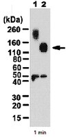
      Molecular Weight~130kDa observed. UniProt describes 2 isoforms produced by alternative splicing at ~106 kDa (Isoform 1), ~107 kDa (Isoform 2). An uncharacterized band may be observed at ~48 kDa in some tissue lysates.
      Physicochemical Information
      Dimensions
      Materials Information
      Toxicological Information
      Safety Information according to GHS
      Safety Information
      Product Usage Statements
      Quality AssuranceEvaluated by Western Blot in untreated and PNGase F treated mouse P7 brain tissue lysate.

      Western Blot Analysis: 0.5 µg/mL of this antibody detected MDGA1 in 5 µg of untreated and PNGase F treated mouse P7 brain tissue lysate. This antibody recognizes glycosolated (>160 kDa) (lane 1) and deglycosolated MDGA1 (lane 2).
      Usage Statement
      • Unless otherwise stated in our catalog or other company documentation accompanying the product(s), our products are intended for research use only and are not to be used for any other purpose, which includes but is not limited to, unauthorized commercial uses, in vitro diagnostic uses, ex vivo or in vivo therapeutic uses or any type of consumption or application to humans or animals.
      Storage and Shipping Information
      Storage ConditionsStable for 1 year at 2-8°C from date of receipt.
      Packaging Information
      Material Size100 µg
      Transport Information
      Supplemental Information
      Specifications
      Global Trade Item Number
      カタログ番号 GTIN
      AB2226 04053252414015

      Documentation

      Anti-MDGA1 Antibody (M)SDS

      タイトル

      英語版製品安全データシート((M)SDS) 

      Anti-MDGA1 Antibody 試験成績書(CoA)

      タイトルロット番号
      Anti-MDGA1 - 2420585 2420585
      Anti-MDGA1 - 2366009 2366009
      Anti-MDGA1 - 3380296 3380296
      Anti-MDGA1 - 3780153 3780153
      Anti-MDGA1 - 3833695 3833695
      Anti-MDGA1 - 3933674 3933674
      Anti-MDGA1 - Q1988383 Q1988383
      Anti-MDGA1 -2820751 2820751
      Anti-MDGA1 -2823633 2823633
      Anti-MDGA1 Polyclonal Antibody 2945401