Monoclonal antibody 76F distinguishes IA-2 from IA-2beta and overlaps an autoantibody epitope.
Piquer, Sandra, et al.
J. Autoimmun., 26: 215-22 (2006)
2006
概要を表示する
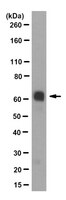
IA-2 and IA-2beta are highly related proteins that are autoantigens in type 1 diabetes, and provide a model for developing reagents and assays that distinguish similar proteins with unique autoantibody epitopes. Monoclonal antibodies (mAb) to IA-2 and IA-2beta were prepared and tested for their ability to bind to the related proteins and their ability to compete for specific autoantibody epitope binding by sera from patients with type 1 diabetes. Monoclonal antibodies that specifically bound IA-2 (76F) or bound both IA-2 and IA-2beta (A9) were isolated and characterized. 76F mAb recognized IA-2 of human, rat and mouse origin in native and denatured forms and had an epitope specificity for residues 626-630 (FEYQD) which are found in the juxtamembrane (JM) region of human and mouse IA-2, but not IA-2beta. This region overlaps with the autoantibody epitope JM2. Binding to the 76F monoclonal antibody was specifically inhibited by sera with antibodies to the JM2 epitope but not with antibodies to the adjacent JM1 epitope, indicating that unique epitopes can be distinguished by this approach. 76F mAb has the unique property to distinguish between the two closely related autoantigens IA-2 and IA-2beta by targeting an IA-2 specific epitope of the juxtamembrane region. The findings define an approach to develop assays for specific antibody epitope measurements which may be relevant for disease prognosis and monitoring intervention therapies. | 16503116
 |