Millipore Sigma Vibrant Logo

CBA001 InnoZyme™ Cathepsin B Activity Assay Kit, Fluorogenic

View This Product on Sigma-Aldrich
CBA001
View Pricing & Availability

Overview

Replacement Information

Key Spec Table

Detection Methods
Fluorometric

Pricing & Availability

Catalogue Number AvailabilityPackaging Qty/Pack Price Quantity
CBA001-1KITCN
Retrieving availability...
Limited Availability
In Stock 
Discontinued
Limited Quantities Available
Availability to be confirmed
    Remaining : Will advise
      Remaining : Will advise
      Will advise
      Contact Customer Service
      Contact Customer Service

      Glass bottle 1 kit
      Retrieving price...
      Price could not be retrieved
      Minimum Quantity is a multiple of
      Maximum Quantity is
      Upon Order Completion More Information
      You Saved ()
       
      Request Pricing
      Description
      OverviewA sensitive fluorogenic assay for the measurement of cathepsin B activity (Excitation max: ~360-380 nm; Emission max: ~430-460 nm). Cathepsin B is a cysteine proteinase which has been associated with increased invasivessness and development of the malignant cell phenotype. Cathepsin B has also been implicated in inflammatory airway disease.
      Catalogue NumberCBA001
      Brand Family Calbiochem®
      Application Data
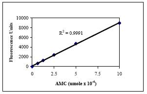
      Calibration curve generated by various dilutions of the free AMC standard included in the kit (above).
      Materials Required but Not Delivered Pipettors or multi-channel pipettors calibrated to target volume
      37°C incubator
      Microplate reader capable of measuring fluorescence at wavelengths 360-380 nm for excitation and 440-460 nm for emission
      Ice bucket
      References
      ReferencesBervar, A., et al. 2003. Biol. Chem. 384, 447.
      Grigolo, B. et al. 2003. Biomaterials 24, 1751.
      Scolaris, A., et al. 2002. Biol. Chem. 383, 1297.
      Murata, M., et al. 1991. FEBS Lett. 280, 307.
      Berquin, I.M. and Sloane, B.F. 1996. Adv. Exp. Med. Biol. 389, 281.
      Burnett, D., 1995. Arch. Biochem. Biophys. 317, 305.
      Reddy, V.Y., et al. 1995. Proc. Natl. Acad. Sci. 92, 3849.
      Buttle, D.J., 1994. In: Immunopharmacology of Joints and Connective Tissue (Dingle, J.T. and Davies, M.E., eds) London: Academic Press. 225.
      Barrett, A.J. and Kirschke, H., 1981. Methods Enzymol. 80, 535.
      Product Information
      Detection methodFluorometric
      Form96 Tests
      Format96-well plate
      Kit containsCathepsin B Enzyme, Calibration Standard, Cathepsin B Substrate, Assay Buffer, Cathepsin B Inhibitor, Reduction Reagent, Cell Lysis Buffer, Microtiter Plate, Plate Sealer, and a user protocol.
      Positive controlCathepsin B
      Quality LevelMQ100
      Applications
      Biological Information
      Assay range0.09 - 100 ng/ml
      Assay time30 min
      Sample TypeCells and tissues
      Physicochemical Information
      Sensitivity0.63 ng/ml
      Dimensions
      Materials Information
      Toxicological Information
      Safety Information according to GHS
      Safety Information
      Product Usage Statements
      Intended useThe Calbiochem® Cathepsin B Activity Assay Kit, Fluorogenic is designed to measure cathepsin B activity in tissues extracts and cell lysates, and for screening cathepsin B inhibitors.

      Note: Following initial use the control Cathepsin B should be dispensed into aliquots and stored at -20°C. Avoid freeze/thaw cycles
      Storage and Shipping Information
      Ship Code Dry Ice Only
      Toxicity Multiple Toxicity Values, refer to MSDS
      Storage Multiple storage requirements
      Storage ConditionsStore the unopened components at -20°C and -70°C as indicated on the labels. All kit components, once opened, can be stored under the following conditions:

      Assay Buffer, Cell Lysis Buffer, 96-Well Plate, and Plate Sealers at Room Temperature

      Cathepsin B Inhibitor, Reduction Reagent, Cathepsin B Substrate, and Calibration Standard at -20°C

      Control Cathepsin B(aliquot) at -70°C
      Protect from Light Protect from light
      Avoid freeze/thaw Avoid freeze/thaw
      Do not freeze Ok to freeze
      Packaging Information
      Transport Information
      Supplemental Information
      Kit containsCathepsin B Enzyme, Calibration Standard, Cathepsin B Substrate, Assay Buffer, Cathepsin B Inhibitor, Reduction Reagent, Cell Lysis Buffer, Microtiter Plate, Plate Sealer, and a user protocol.
      Specifications
      Global Trade Item Number
      Catalogue Number GTIN
      CBA001-1KITCN 04055977220810

      Documentation

      InnoZyme™ Cathepsin B Activity Assay Kit, Fluorogenic SDS

      Title

      Safety Data Sheet (SDS) 

      InnoZyme™ Cathepsin B Activity Assay Kit, Fluorogenic Certificates of Analysis

      TitleLot Number
      CBA001

      References

      Reference overview
      Bervar, A., et al. 2003. Biol. Chem. 384, 447.
      Grigolo, B. et al. 2003. Biomaterials 24, 1751.
      Scolaris, A., et al. 2002. Biol. Chem. 383, 1297.
      Murata, M., et al. 1991. FEBS Lett. 280, 307.
      Berquin, I.M. and Sloane, B.F. 1996. Adv. Exp. Med. Biol. 389, 281.
      Burnett, D., 1995. Arch. Biochem. Biophys. 317, 305.
      Reddy, V.Y., et al. 1995. Proc. Natl. Acad. Sci. 92, 3849.
      Buttle, D.J., 1994. In: Immunopharmacology of Joints and Connective Tissue (Dingle, J.T. and Davies, M.E., eds) London: Academic Press. 225.
      Barrett, A.J. and Kirschke, H., 1981. Methods Enzymol. 80, 535.
      User Protocol

      Revision05-June-2012 JSW
      Form96 Tests
      Format96-well plate
      Detection methodFluorometric
      StorageStore the unopened components at -20°C and -70°C as indicated on the labels. All kit components, once opened, can be stored under the following conditions:

      Assay Buffer, Cell Lysis Buffer, 96-Well Plate, and Plate Sealers at Room Temperature

      Cathepsin B Inhibitor, Reduction Reagent, Cathepsin B Substrate, and Calibration Standard at -20°C

      Control Cathepsin B(aliquot) at -70°C
      Intended useThe Calbiochem® Cathepsin B Activity Assay Kit, Fluorogenic is designed to measure cathepsin B activity in tissues extracts and cell lysates, and for screening cathepsin B inhibitors.

      Note: Following initial use the control Cathepsin B should be dispensed into aliquots and stored at -20°C. Avoid freeze/thaw cycles
      BackgroundExtensive studies have demonstrated increased cathepsin B levels in human tumors suggesting a role in invasion and metastasis. Although lysosomal localization of the enzyme suggests it functions primarily as a component of the protein degradation system, recent data have pointed to alternative localization of cathepsin B including the nucleus, the cytoplasm, and the plasma membrane. Additionally, there is evidence of the presence of the active enzyme in the extracellular matrix suggesting "escape" of the active enzyme from lysosome or extracellular mechanism for proenzyme activation. Extracellular cathepsin B has been implicated in inflammatory airway disease and in bone and joint disorders. Cathepsin B has been successfully used as a marker to monitor the differentiation process in engineered cartilage.
      Principles of the assayThe InnoZyme Cathepsin B Activity Assay Kit, Fluorogenic is designed for the quantitative in vitro determination of cathepsin B activity in a 96-well format. The test utilizes the ability of cathepsin B to digest the synthetic substrate Z-Arg-Arg AMC. Released free AMC is determined fluorometrically at excitation wavelength 360-380 nm and emission wavelength 440-460 nm. The activity of the cathepsin B can be quantified with an AMC standard, or can be displayed as fluorescence units (FU). For biological samples the cathepsin B inhibitor, CA-074, can be used as a negative control.
      Materials provided• Cathepsin B Substrate (Kit Component No. JA7740-40UL): 1 vial, 40 µl of Z-Arg-Arg AMC supplied as a 100-fold concentrated solution in DMSO
      • Control Cathepsin B (Kit Component No. JA7741-20UL): 1 vial, 20 µl of affinity purified, native cathepsin B, manufactured by EMD Biosciences
      • Calibration Standard (Kit Component No. JA7742-100UL): 1 vial, 100 µl, of 2 mM 7-Amino-4-methylcoumarin (AMC) in DMSO supplied as a 100-fold concentrated solution
      • Cathepsin B Inhibitor (Kit Component No. JA7743-40UL): 1 vial, 40 µl of CA-074 provided as a 10-fold concentrated solution
      • Reduction Reagent (Kit Component No. JA7744-80UL): 1 vial, 80 µl of 1 M cysteine provided as a 250-fold concentrated solution
      • Assay Buffer (Kit Component No. JA7745-20ML): 1 bottle, 20 ml of MES buffer, pH 6.0, containing EDTA.
      • Cell Lysis Buffer (Cat. No.71009): 1 bottle, 10 ml of CytoBuster™ Protein Extraction Reagent
      • 96-Well Plate (Kit Component No. JA7666-1EA): 1 each
      • Plate Sealer (Kit Component No. JB155-EA): 2 each
      Materials Required but not provided Pipettors or multi-channel pipettors calibrated to target volume
      37°C incubator
      Microplate reader capable of measuring fluorescence at wavelengths 360-380 nm for excitation and 440-460 nm for emission
      Ice bucket
      PreparationDilute samples with dilution buffer as necessary. Note: if the measured FU exceeds 9,000 the sample should be diluted further. • Cell lysate: wash cell pellet with ice-cold PBS. Add 500-1000 µl of cell lysis buffer (~1 ml per 1x10⁷ cells) and incubate on ice for 30 min. Vortex and centrifuge the lysate at 14,000 x g in a pre-cooled tabletop microcentrifuge. Immediately transfer the supernatant to a fresh microcentrifuge tube and discard the pellet. Dilute the lysate 1:5 or 1:10 before determining the protein concentration via BCA protein assay.
      Reagent preparation• Dilution Buffer: dilute assay buffer 1:10 with dH2O Example: add 0.5 ml of assay buffer to 4.5 ml of distilled water. • Standard: the free AMC standard is supplied as a 2 mM stock (100-fold concentrated) solution. To create a calibration curve prepare serial dilutions of the standard ranging from a concentration of 20 µM to a concentration of 1.25 µM. Example: label 6 eppendorf tubes. Pipet 495 µl of the dilution buffer into the first tube and 250 µl into the remaining tubes. Add 5 µl of the 2 mM AMC standard to the first tube. Vortex and transfer 250 µl from this tube to the next tube. Continue serial dilutions by transferring 250 µl up to tube #5. The tube labeled #6 will contain dilution buffer only (blank). • Substrate Working Solution: substrate should be diluted 100 times with distilled water. Example: to prepare 2 ml of substrate solution add 20 µl of the substrate stock solution to 1.98 ml of distilled water. • Activation Buffer: cysteine is supplied as a 250-fold concentrated reagent. To prepare activation buffer 8 µl of 1 M cysteine should be added to 2 ml of the supplied assay buffer (this is enough for half of the plate). • Inhibitor: CA-074 inhibitor is supplied as a 10-fold stock solution and should be diluted with activation buffer. Example: dilute 10 µl of the stock solution with 90 µl of the activation buffer to yield 100 µl of working solution. • Control: dilute the control human cathepsin B with dilution buffer as directed on the vial label. The cathepsin B enzyme should be dispensed into aliquots and stored at -70°C to maintain activity.
      Detailed protocolNote: All reagents necessary to perform the assay are supplied with this kit. Warm all components to 15-25°C before use. Control and samples should be kept at -70°C until just prior to use. It is recommended that all standards, controls, and samples be assayed in duplicate.

      1. Remove the desired number of strips from the plate and return the unused strips to the foil pouch.
      2. Add 25 µl Activation Buffer to each well. Note: to inactivate cathepsin B activity use 20 µl of activation buffer and 5 µl of inhibitor.
      3. Add 50 µl of standard, control, or sample to individual, designated wells. Blank should contain 50 µl of dilution buffer. Note: all biological samples should be run in parallel with inhibitor (and activation buffer) and without inhibitor.
      4. Pre-incubate the plate at room temperature for 5 min.
      5. Add 25 µl of substrate solution.
      6. Seal the plate tightly and incubate at 37°C for 30 min.
      7. Read the fluorescence of free AMC on a fluorescence plate reader at excitation wavelength 360-380 nm and emission wavelength 440-460 nm.
      CalculationsThe data obtained can be displayed in two ways: 1. As fluorescence units: correct the fluorescence value of all samples by subtracting the value of the blank, and then calculate the mean fluorescence value for each sample in duplicate. For the biological samples also subtract the value of the sample assayed with inhibitor. OR 2. As µmole of free AMC/mg total protein/time (min.): calculate the mean fluorescence value as stated above. Plot a graph correlating the mean fluorescence values of the AMC standards (y-axis) to their concentration in µmole (x-axis). The cathepsin B activity of the unknown samples can be interpolated from the standard curve. Calculate the amount (µmol) of free AMC per mg of total protein and time unit.
      Standard curve

      Figure 1: Standard Curve

      Calibration curve generated by various dilutions of the free AMC standard included in the kit (above).

      Example data

      Figure 2: Native cathepsin B activity

      The activity of native cathepsin B (Cat. No. 219364) with Z-Arg-Arg AMC substrate in MES buffer, pH 6.0, in the presence of EDTA and cysteine. After incubation at 37°C for 30 min the free AMC was measured at excitation 360 nm and emission 460 nm (above).

      Table 1: Cathepsin B activity in human tissue extracts

      Cathepsin B activity in human tissue extracts as measured with Cathepsin B Activity Assay kit (above).

      Table 2: Cathepsin B activity in cell lysates

      Cathepsin B activity in five human cell line lysates as measured with Cathepsin B Activity Assay kit. Cell lysis buffer (CytoBuster™ Protein Extraction Reagent, Cat. No. 71009) was used to prepare lysates (above).

      Sensitivity0.63 ng/ml
      Sensitivity NotesThe minimum detectable activity is determined to be 0.63 ng/ml. This was calculated based on the concentration of cathepsin B at two standard deviations above the mean fluorescence unit values of ten blank readings. Affinity purified, native human cathepsin B (EMD Biosciences, Cat. No. 219362) was used to determine the sensitivity of the assay
      Assay Range0.09 - 100 ng/ml
      Specificity

      Table 3: Cross reactivity with others cathepsins

      Registered TrademarksCalbiochem® is a registered trademark of EMD Chemicals, Inc.
      CytoBuster™, InteractivePathways™ and InnoZyme™ are trademarks of EMD Chemicals, Inc.