Millipore Sigma Vibrant Logo

MABN1847 Anti-Synapsin-1 Antibody, clone MEGS3-10A

View This Product on Sigma-Aldrich
MABN1847
100 µL  
Retrieving price...
Price could not be retrieved
Minimum Quantity is a multiple of
Maximum Quantity is
Upon Order Completion More Information
You Saved ()
 
Request Pricing
Limited Availability
In Stock 
Discontinued
Limited Quantities Available
Availability to be confirmed
    Remaining : Will advise
      Remaining : Will advise
      Will advise
      Contact Customer Service
      Contact Customer Service

      Special Offers


      Contact Customer Service

      Overview

      Replacement Information

      Key Spec Table

      Species ReactivityKey ApplicationsHostFormatAntibody Type
      MWB, ICCMPurifiedMonoclonal Antibody
      Description
      Catalogue NumberMABN1847
      DescriptionAnti-Synapsin-1 Antibody, clone MEGS3-10A
      Alternate Names
      • Synapsin-1
      • Synapsin I
      Background InformationSynapsin-1 (UniProt O88935; also known as Synapsin I) is encoded by the Syn1 (also known as Syn-1) gene (Gene ID 20964) in murine species. The synapins constitute a family of abundant neuronal phosphoproteins that regulate SV trafficking and neurotransmitter release at the pre-synaptic terminal. Three genes exisit in mammals, encoding altogether 11 synapsin members by alternative splicings (Synapsin Ia & Ib by SYN1, Synapsin IIa & IIb by SYN2, Synapsin IIIa through IIIf by SYN3), Syn III is the most precociously expressed isoform that has a role in the early phases of neural development and is downregulated in mature neurons. On the other hand, Syn I and Syn II are expressed at low levels at birth and their expressions progressively increase during synaptogenesis to reach a stable plateau at 1–2 months of life. The NH2-terminal region is divided in A, B, and C domains and is highly conserved among synapsin isoforms. Synapsins are regulated by postransloational phosphorylations. Domain A contains PKA and CaMKI/IV phosphorylation sites, domain B contains MAPK/Erk phosphorylation sites, and domain C is phosphorylated by Src. The c-terminal regions contain spliced domains and diverge among synapsin isoforms (domain D in Syn Ia and Ib, domain G in Syn IIa and IIb, domain H in Syn IIa, and domain J in Syn IIIa), although they all bear proline-rich regions binding to several SH3-containing proteins and additional phosphorylation sites for CaMKII, MAPK/Erk, and cdk1/5.
      References
      Product Information
      FormatPurified
      PresentationPurified mouse monoclonal IgG2bκ antibody in buffer containing 0.1 M Tris-Glycine (pH 7.4), 150 mM NaCl with 0.05% sodium azide.
      Quality LevelMQ100
      Applications
      Key Applications
      • Western Blotting
      • Immunocytochemistry
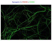
      Application NotesImmunocytochemistry Analysis: The ascites prior to antibody purification detected Synapsin-1 in primary mouse cortical neurons (Courtesy of Professor Michael E. Greenberg, Harvard Medical School, Boston, MA).
      Western Blotting Analysis: An 1:1000 dilution of the ascites prior to antibody purification detected a higher Synapsin-1 expression in brain lysate from a 60-day old (P60) mouse than from a 6-day old (P6) mouse (Courtesy of Professor Michael E. Greenberg, Harvard Medical School, Boston, MA).
      Biological Information
      ImmunogenRecombinant full-length mouse synapsin-1.
      CloneMEGS3-10A
      ConcentrationPlease refer to lot specific datasheet.
      HostMouse
      IsotypeIgMκ
      Species Reactivity
      • Mouse
      Antibody TypeMonoclonal Antibody
      Entrez Gene Number
      Gene Symbol
      • Syn1
      • Syn-1
      UniProt Number
      Molecular Weight~75 kDa observed.
      Physicochemical Information
      Dimensions
      Materials Information
      Toxicological Information
      Safety Information according to GHS
      Safety Information
      Product Usage Statements
      Quality AssuranceEvaluated by Western Blotting in mouse brain tissue lysate.

      Western Blotting Analysis: 1.0 µg/mL of this antibody detected Synapsin-1 in 10 µg of mouse brain tissue lysate.
      Usage Statement
      • Unless otherwise stated in our catalog or other company documentation accompanying the product(s), our products are intended for research use only and are not to be used for any other purpose, which includes but is not limited to, unauthorized commercial uses, in vitro diagnostic uses, ex vivo or in vivo therapeutic uses or any type of consumption or application to humans or animals.
      Storage and Shipping Information
      Storage ConditionsStable for 1 year at 2-8°C from date of receipt.
      Packaging Information
      Material Size100 µL
      Transport Information
      Supplemental Information
      Specifications
      Global Trade Item Number
      Catalogue Number GTIN
      MABN1847 04055977311860