Arterial dysfunction but maintained systemic blood pressure in cavin-1-deficient mice.
Swärd, K; Albinsson, S; Rippe, C
PloS one
9
e92428
2014
Mostra il sommario
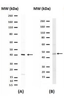
Caveolae are omega-shaped plasma membrane micro-domains that are abundant in cells of the vascular system. Formation of caveolae depends on caveolin-1 and cavin-1 and lack of either protein leads to loss of caveolae. Mice with caveolin-1 deficiency have dysfunctional blood vessels, but whether absence of cavin-1 similarly leads to vascular dysfunction is not known. Here we addressed this hypothesis using small mesenteric arteries from cavin-1-deficient mice. Cavin-1-reporter staining was intense in mesenteric arteries, brain arterioles and elsewhere in the vascular system, with positive staining of both endothelial and smooth muscle cells. Arterial expression of cavin-1, -2 and -3 was reduced in knockout (KO) arteries as was expression of caveolin-1, -2 and -3. Caveolae were absent in the endothelial and smooth muscle layers of small mesenteric arteries as determined by electron microscopy. Arginase, a negative regulator of nitric oxide production, was elevated in cavin-1 deficient arteries as was contraction in response to the α1-adrenergic agonist cirazoline. Detailed assessment of vascular dimensions revealed increased media thickness and reduced distensibility, arguing that enhanced contraction was due to increased muscle mass. Contrasting with increased α1-adrenergic contraction, myogenic tone was essentially absent and this appeared to be due in part to increased nitric oxide production. Vasomotion was less frequent in the knock-out vessels. In keeping with the opposing influences on arterial resistance of increased agonist-induced contractility and reduced myogenic tone, arterial blood pressure was unchanged in vivo. We conclude that deficiency of cavin-1 affects the function of small arteries, but that opposing influences on arterial resistance balance each other such that systemic blood pressure in unstressed mice is well maintained. | Western Blotting | 24658465
 |
High flow conditions increase connexin43 expression in a rat arteriovenous and angioinductive loop model.
Schmidt, VJ; Hilgert, JG; Covi, JM; Weis, C; Wietbrock, JO; de Wit, C; Horch, RE; Kneser, U
PloS one
8
e78782
2013
Mostra il sommario
Gap junctions are involved in vascular growth and their expression pattern is modulated in response to hemodynamic conditions. They are clusters of intercellular channels formed by connexins (Cx) of which four subtypes are expressed in the cardiovascular system, namely Cx37, Cx40, Cx43 and Cx45. We hypothesize that high flow conditions affect vascular expression of Cx in vivo. To test this hypothesis, flow hemodynamics and subsequent changes in vascular expression of Cx were studied in an angioinductive rat arteriovenous (AV) loop model. Fifteen days after interposition of a femoral vein graft between femoral artery and vein encased in a fibrin-filled chamber strong neovascularization was evident that emerged predominantly from the graft. Blood flow through the grafted vessel was enhanced ∼4.5-fold accompanied by increased pulsatility exceeding arterial levels. Whereas Cx43 protein expression in the femoral vein is negligible at physiologic flow conditions as judged by immunostaining its expression was enhanced in the endothelium of the venous graft exposed to these hemodynamic changes for 5 days. This was most likely due to enhanced transcription since Cx43 mRNA increased likewise, whereas Cx37 mRNA expression remained unaffected and Cx40 mRNA was reduced. Although enhanced Cx43 expression in regions of high flow in vivo has already been demonstrated, the arteriovenous graft used in the present study provides a reliable model to verify an association between Cx43 expression and high flow conditions in vivo that was selective for this Cx. We conclude that enhancement of blood flow and its oscillation possibly associated with the transition from laminar to more turbulent flow induces Cx43 expression in a vein serving as an AV loop. It is tempting to speculate that this upregulation is involved in the vessel formation occuring in this model as Cx43 was suggested to be involved in angiogenesis. | | 24236049
 |
Inverse Relationship between Tumor Proliferation Markers and Connexin Expression in a Malignant Cardiac Tumor Originating from Mesenchymal Stem Cell Engineered Tissue in a Rat in vivo Model.
Spath, C; Schlegel, F; Leontyev, S; Mohr, FW; Dhein, S
Frontiers in pharmacology
4
42
2013
Mostra il sommario
Recently, we demonstrated the beneficial effects of engineered heart tissues for the treatment of dilated cardiomyopathy in rats. For further development of this technique we started to produce engineered tissue (ET) from mesenchymal stem cells. Interestingly, we observed a malignant tumor invading the heart with an inverse relationship between proliferation markers and connexin expression.Commercial CD54(+)/CD90(+)/CD34(-)/CD45(-) bone marrow derived mesenchymal rat stem cells (cBM-MSC), characterized were used for production of mesenchymal stem-cell-ET (MSC-ET) by suspending them in a collagen I, matrigel-mixture and cultivating for 14 days with electrical stimulation. Three MSC-ET were implanted around the beating heart of adult rats for days. Another three MSC-ET were produced from freshly isolated rat bone marrow derived stem cells (sBM-MSC).Three weeks after implantation of the MSC-ETs the hearts were surgically excised. While in 5/6 cases the ET was clearly distinguishable and was found as a ring containing mostly connective tissue around the heart, in 1/6 the heart was completely surrounded by a huge, undifferentiated, pleomorphic tumor originating from the cMSC-ET (cBM-MSC), classified as a high grade malignant sarcoma. Quantitatively we found a clear inverse relationship between cardiac connexin expression (Cx43, Cx40, or Cx45) and increased Ki-67 expression (Cx43: p less than 0.0001, Cx45: p less than 0.03, Cx40: p less than 0.014). At the tumor-heart border there were significantly more Ki-67 positive cells (p = 0.001), and only 2% Cx45 and Ki-67-expressing cells, while the other connexins were nearly completely absent (p less than 0.0001). Conclusion and Hypothesis: These observations strongly suggest the hypothesis, that invasive tumor growth is accompanied by reduction in connexins. This implicates that gap junction communication between tumor and normal tissue is reduced or absent, which could mean that growth and differentiation signals can not be exchanged. | | 23616767
 |
Role of connexin-43 in protective PI3K-Akt-GSK-3β signaling in cardiomyocytes.
Ishikawa, S; Kuno, A; Tanno, M; Miki, T; Kouzu, H; Itoh, T; Sato, T; Sunaga, D; Murase, H; Miura, T
American journal of physiology. Heart and circulatory physiology
302
H2536-44
2011
Mostra il sommario
Sarcolemmal connexin-43 (Cx43) and mitochondrial Cx43 play distinct roles: formation of gap junctions and production of reactive oxygen species (ROS) for redox signaling. In this study, we examined the hypothesis that Cx43 contributes to activation of a major cytoprotective signal pathway, phosphoinositide 3-kinase (PI3K)-Akt-glycogen synthase kinase-3β (GSK-3β) signaling, in cardiomyocytes. A δ-opioid receptor agonist {[d-Ala(2),d-Leu(5)]enkephalin acetate (DADLE)}, endothelin-1 (ET-1), and insulin-like growth factor-1 (IGF-1) induced phosphorylation of Akt and GSK-3β in H9c2 cardiomyocytes. Reduction of Cx43 protein to 20% of the normal level by Cx43 small interfering RNA abolished phosphorylation of Akt and GSK-3β induced by DADLE or ET-1 but not that induced by IGF-1. DADLE and IGF-1 protected H9c2 cells from necrosis after treatment with H(2)O(2) or antimycin A. The protection by DADLE or ET-1, but not that by IGF-1, was lost by reduction of Cx43 protein expression. In contrast to Akt and GSK-3β, PKC-ε, ERK and p38 mitogen-activated protein kinase were phosphorylated by ET-1 in Cx43-knocked-down cells. Like diazoxide, an activator of the mitochondrial ATP-sensitive K(+) channel, DADLE and ET-1 induced significant ROS production in mitochondria, although such an effect was not observed for IGF-1. Cx43 knockdown did not attenuate the mitochondrial ROS production by DADLE or ET-1. Cx43 was coimmunoprecipitated with the β-subunit of G protein (Gβ), and knockdown of Gβ mimicked the effect of Cx43 knockdown on ET-1-induced phosphorylation of Akt and GSK-3β. These results suggest that Cx43 contributes to activation of class I(B) PI3K in PI3K-Akt-GSK-3β signaling possibly as a cofactor of Gβ in cardiomyocytes. | | 22505645
 |
Role of connexin40 in the autoregulatory response of the afferent arteriole.
Sorensen, CM; Giese, I; Braunstein, TH; Brasen, JC; Salomonsson, M; Holstein-Rathlou, NH
American journal of physiology. Renal physiology
303
F855-63
2011
Mostra il sommario
Connexins in renal arterioles affect autoregulation of arteriolar tonus and renal blood flow and are believed to be involved in the transmission of the tubuloglomerular feedback (TGF) response across the cells of the juxtaglomerular apparatus. Connexin40 (Cx40) also plays a significant role in the regulation of renin secretion. We investigated the effect of deleting the Cx40 gene on autoregulation of afferent arteriolar diameter in response to acute changes in renal perfusion pressure. The experiments were performed using the isolated blood perfused juxtamedullary nephron preparation in kidneys obtained from wild-type or Cx40 knockout mice. Renal perfusion pressure was increased in steps from 75 to 155 mmHg, and the response in afferent arteriolar diameter was measured. Hereafter, a papillectomy was performed to inhibit TGF, and the pressure steps were repeated. Conduction of intercellular Ca(2+) changes in response to local electrical stimulation was examined in isolated interlobular arteries and afferent arterioles from wild-type or Cx40 knockout mice. Cx40 knockout mice had an impaired autoregulatory response to acute changes in renal perfusion pressure compared with wild-type mice. Inhibition of TGF by papillectomy significantly reduced autoregulation of afferent arteriolar diameter in wild-type mice. In Cx40 knockout mice, papillectomy did not affect the autoregulatory response, indicating that these mice have no functional TGF. Also, Cx40 knockout mice showed no conduction of intercellular Ca(2+) changes in response to local electrical stimulation of interlobular arteries, whereas the Ca(2+) response to norepinephrine was unaffected. These results suggest that Cx40 plays a significant role in the renal autoregulatory response of preglomerular resistance vessels. | | 22811484
 |
GM-CSF causes a Paradoxical Increase in the BH3-only Pro-Apoptotic Protien BIM in Human Neutrophils.
Cowburn AS, Summers C, Dunmore BJ, Farahi N, Hayhoe RP, Print CG, Cook SJ, Chilvers ER
Am J Respir Cell Mol Biol
2009
Mostra il sommario
Neutrophil apoptosis is essential for the resolution of inflammation but delayed by several inflammatory mediators. In such terminally differentiated cells it has been uncertain whether these agents can inhibit apoptosis through transcriptional regulation of anti-death (Bcl-XL, Mcl-1, Bcl2A1) or BH3-only (Bim, Bid, Puma) Bcl2-family proteins. We report that GM-CSF and TNFa prevent the normal time-dependent loss of Mcl-1 and Bcl2A1 in neutrophils and demonstrate that they cause a NF-kB-dependent increase in Bcl-XL transcription/translation. Surprisingly, we show that GM-CSF and TNFa increase and/or maintain mRNA levels for the pro-apoptotic BH3-only protein Bid and that GM-CSF has a similar NF-kB-dependent effect on Bim transcription and BimEL expression. The in-vivo relevance of these findings was shown by the demonstration that GM-CSF is the dominant neutrophil survival factor present in lung lavage from patients with ventilator-associated pneumonia and confirmation of an increase lung neutrophil Bim mRNA. Finally GM-CSF caused mitochondrial location of Bim and a switch in phenotype to a cell that displays accelerated caspase-9-dependent apoptosis. This study demonstrates the capacity of neutrophil survival agents to induce a paradoxical increase in the pro-apoptotic proteins Bid and Bim and suggests that this may function to facilitate rapid apoptosis at the termination of the inflammatory cycle. | | 20705940
 |
Loss of connexin40 is associated with decreased endothelium-dependent relaxations and eNOS levels in the mouse aorta.
Alonso, F; Boittin, FX; Bény, JL; Haefliger, JA
American journal of physiology. Heart and circulatory physiology
299
H1365-73
2009
Mostra il sommario
Upon agonist stimulation, endothelial cells trigger smooth muscle relaxation through the release of relaxing factors such as nitric oxide (NO). Endothelial cells of mouse aorta are interconnected by gap junctions made of connexin40 (Cx40) and connexin37 (Cx37), allowing the exchange of signaling molecules to coordinate their activity. Wild-type (Cx40(+/+)) and hypertensive Cx40-deficient mice (Cx40(-/-)), which also exhibit a marked decrease of Cx37 in the endothelium, were used to investigate the link between the expression of endothelial connexins (Cx40 and Cx37) and endothelial nitric oxide synthase (eNOS) expression and function in the mouse aorta. With the use of isometric tension measurements in aortic rings precontracted with U-46619, a stable thromboxane A(2) mimetic, we first demonstrate that ACh- and ATP-induced endothelium-dependent relaxations solely depend on NO release in both Cx40(+/+) and Cx40(-/-) mice, but are markedly weaker in Cx40(-/-) mice. Consistently, both basal and ACh- or ATP-induced NO production were decreased in the aorta of Cx40(-/-) mice. Altered relaxations and NO release from aorta of Cx40(-/-) mice were associated with lower expression levels of eNOS in the aortic endothelium of Cx40(-/-) mice. Using immunoprecipitation and in situ ligation assay, we further demonstrate that eNOS, Cx40, and Cx37 tightly interact with each other at intercellular junctions in the aortic endothelium of Cx40(+/+) mice, suggesting that the absence of Cx40 in association with altered Cx37 levels in endothelial cells from Cx40(-/-) mice participate to the decreased levels of eNOS. Altogether, our data suggest that the endothelial connexins may participate in the control of eNOS expression levels and function. | | 20802140
 |
An angiotensin II- and NF-kappaB-dependent mechanism increases connexin 43 in murine arteries targeted by renin-dependent hypertension.
Alonso, F; Krattinger, N; Mazzolai, L; Simon, A; Waeber, G; Meda, P; Haefliger, JA
Cardiovascular research
87
166-76
2009
Mostra il sommario
Connexins (Cxs) play a role in the contractility of the aorta wall. We investigated how connexins of the endothelial cells (ECs; Cx37, Cx40) and smooth muscle cells (SMCs; Cx43, Cx45) of the aorta change during renin-dependent and -independent hypertension.We subjected both wild-type (WT) mice and mice lacking Cx40 (Cx40(-/-)), to either a two-kidney, one-clip procedure or to N-nitro-l-arginine-methyl-ester treatment, which induce renin-dependent and -independent hypertension, respectively. All hypertensive mice featured a thickened aortic wall, increased levels of Cx37 and Cx45 in SMC, and of Cx40 in EC (except in Cx40(-/-) mice). Cx43 was up-regulated, with no effect on its S368 phosphorylation, only in the SMCs of renin-dependent models of hypertension. Blockade of the renin-angiotensin system of Cx40(-/-) mice normalized blood pressure and prevented both aortic thickening and Cx alterations. Ex vivo exposure of WT aortas, carotids, and mesenteric arteries to physiologically relevant levels of angiotensin II (AngII) increased the levels of Cx43, but not of other Cx. In the aortic SMC line of A7r5 cells, AngII activated kinase-dependent pathways and induced binding of the nuclear factor-kappa B (NF-kappaB) to the Cx43 gene promoter, increasing Cx43 expression.In both large and small arteries, hypertension differently regulates Cx expression in SMC and EC layers. Cx43 is selectively increased in renin-dependent hypertension via an AngII activation of the extracellular signal-regulated kinase and NF-kappaB pathways. Testo completo dell'articolo | | 20110337
 |
Role of caveolar compartmentation in endothelium-derived hyperpolarizing factor-mediated relaxation: Ca2+ signals and gap junction function are regulated by caveolin in endothelial cells.
Saliez, J; Bouzin, C; Rath, G; Ghisdal, P; Desjardins, F; Rezzani, R; Rodella, LF; Vriens, J; Nilius, B; Feron, O; Balligand, JL; Dessy, C
Circulation
117
1065-74
2008
Mostra il sommario
In endothelial cells, caveolin-1, the structural protein of caveolae, acts as a scaffolding protein to cluster lipids and signaling molecules within caveolae and, in some instances, regulates the activity of proteins targeted to caveolae. Specifically, different putative mediators of the endothelium-derived hyperpolarizing factor (EDHF)-mediated relaxation are located in caveolae and/or regulated by the structural protein caveolin-1, such as potassium channels, calcium regulatory proteins, and connexin 43, a molecular component of gap junctions.Comparing relaxation in vessels from caveolin-1 knockout mice and their wild-type littermates, we observed a complete absence of EDHF-mediated vasodilation in isolated mesenteric arteries from caveolin-1 knockout mice. The absence of caveolin-1 is associated with an impairment of calcium homeostasis in endothelial cells, notably, a decreased activity of Ca2+-permeable TRPV4 cation channels that participate in nitric oxide- and EDHF-mediated relaxation. Moreover, morphological characterization of caveolin-1 knockout and wild-type arteries showed fewer gap junctions in vessels from knockout animals associated with a lower expression of connexins 37, 40, and 43 and altered myoendothelial communication. Finally, we showed that TRPV4 channels and connexins colocalize with caveolin-1 in the caveolar compartment of the plasma membrane.We demonstrated that expression of caveolin-1 is required for EDHF-related relaxation by modulating membrane location and activity of TRPV4 channels and connexins, which are both implicated at different steps in the EDHF-signaling pathway. | | 18268148
 |
Connexin isoform expression in smooth muscle cells and endothelial cells of hamster cheek pouch arterioles and retractor feed arteries.
Hakim, CH; Jackson, WF; Segal, SS
Microcirculation (New York, N.Y. : 1994)
15
503-14
2008
Mostra il sommario
Gap junction channels formed by connexin (Cx) protein subunits enable cell-to-cell conduction of vasoactive signals. Given the lack of quantitative measurements of Cx expression in microvascular endothelial cells (EC) and smooth muscle cells (SMC), the objective was to determine whether Cx expression differed between EC and SMC of resistance microvessels for which conduction is well-characterized.Cheek pouch arterioles (CPA) and retractor feed arteries (RFA) were hand-dissected and dissociated to obtain SMC or endothelial tubes. In complementary experiments, small intestine was dissociated to obtain SMC. Following reverse transcription, quantitative Real-Time Polymerase Chain Reaction (qRT-PCR) was performed by using specific primers and fluorescent probes for Cx37, Cx40, and Cx43. Smooth muscle alpha-actin (SMAA) and platelet endothelial cell adhesion molecule-1 (PECAM-1) served as respective reference genes.Transcript copy numbers were similar for each Cx isoform in EC from CPA and RFA (approximately 0.5 Cx/PECAM-1). For SMC, Cx43 transcript in CPA and RFA (less than 0.1 Cx/SMAA) was less (p less than 0.05) than that in small intestine (approximately 0.4 Cx/SMAA). Transcripts for Cx37 and Cx40 were also detected in SMC. Punctate immunolabeling for each Cx isoform was pronounced at EC borders and that for Cx43 was pronounced in SMC of small intestine. In contrast, Cx immunolabeling was not detected in SMC of CPA or RFA.Connexin expression occurs primarily within the endothelium of arterioles and feed arteries, supporting a highly effective pathway for conducting vasoactive signals along resistance networks. The apparent paucity of Cx expression within SMC underscores discrete homocellular coupling and focal localization of myoendothelial gap junctions. | | 19086260
 |


