Millipore Sigma Vibrant Logo
Attenzione: Ci siamo trasferiti. Non è più possibile acquistare i prodotti Merck Millipore nel sito MerckMillipore.comPer saperne di più

AB746P Anti-Collagen Type II Antibody

AB746P
100 µg  
Purchase on Sigma-Aldrich

Offerte speciali

Panoramica

Replacement Information

Offerte speciali

Tabella delle specifiche principali

Species ReactivityKey ApplicationsHostFormatAntibody Type
BELISA, RIA, IHC, IH(P)RbPurifiedPolyclonal Antibody
Description
Catalogue NumberAB746P
ReplacesAB746
Brand Family Chemicon®
Trade Name
  • Chemicon
DescriptionAnti-Collagen Type II Antibody
References
Product Information
FormatPurified
PresentationProtein G affinity purified fraction. Liquid in 0.1M phosphate, 0.09M sodium chloride, pH 7.2
Quality LevelMQ100
Applications
ApplicationDetect Collagen Type II using this Anti-Collagen Type II Antibody validated for use in ELISA, RIA, IH, IH(P).
Key Applications
  • ELISA
  • Radioimmunoassay
  • Immunohistochemistry
  • Immunohistochemistry (Paraffin)
Applications Not Recommended
  • Western Blotting
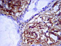
Application NotesImmunohistochemistry on fresh frozen or lightly fixed in 2% PFA, or acetone-fixed specimens: 1:40. Bovine articular cartilage tissue recommended as positive control.

Immunohistochemistry on 2% formalin- or acetone-fixed, paraffin-embedded tissues requires HIER and enhanced enzymatic detection.

Radioimmunoassay

ELISA

Not recommended for western blots.

Optimal working dilutions must be determined by the end user.
Biological Information
ConcentrationPlease refer to the Certificate of Analysis for the lot-specific concentration.
HostRabbit
SpecificityReacts with bovine collagen type II protein. Antibody shows less than 0.1% reactivity with bovine Collagen Types I, IX and less than 3% reactivity with bovine Collagen Type XI by RIA. Cross reactivity with other ECM proteins untested.
Species Reactivity
  • Bovine
Species Reactivity NoteImmunohistochemistry: Fresh frozen or lightly fixed in 2% paraformaldehyde or acetone: 1:40; 2% formalin- or acetone-fixed paraffin-embedded tissues require HIER and enhanced enzymatic detection.

ELISA

RIA

Not recommended for western blots

Optimal working dilutions must be determined by end user.
Antibody TypePolyclonal Antibody
Entrez Gene Number
Entrez Gene SummaryThis gene encodes the alpha-1 chain of type II collagen, a fibrillar collagen found in cartilage and the vitreous humor of the eye. Mutations in this gene are associated with achondrogenesis, chondrodysplasia, early onset familial osteoarthritis, SED congenita, Langer-Saldino achondrogenesis, Kniest dysplasia, Stickler syndrome type I, and spondyloepimetaphyseal dysplasia Strudwick type. In addition, defects in processing chondrocalcin, a calcium binding protein that is the C-propeptide of this collagen molecule, are also associated with chondrodysplasia. There are two transcripts identified for this gene.
Gene Symbol
  • COL2A1
  • SEDC
  • COL11A3
  • ANFH
  • MGC131516
  • chondrocalcin
  • AOM
UniProt Number
UniProt SummaryFUNCTION: SwissProt: P02458 # Type II collagen is specific for cartilaginous tissues. It is essential for the normal embryonic development of the skeleton, for linear growth and for the ability of cartilage to resist compressive forces.
SIZE: 1487 amino acids; 141785 Da
SUBUNIT: Homotrimers of alpha 1(II) chains.
SUBCELLULAR LOCATION: Secreted, extracellular space, extracellular matrix (By similarity).
TISSUE SPECIFICITY: High expression of isoform 2 in juvenile chondrocyte and low in fetal chondrocyte.
PTM: Prolines at the third position of the tripeptide repeating unit (G-X-Y) are hydroxylated in some or all of the chains. & The N-telopeptide is covalently linked to the helical COL2 region of alpha 1(IX), alpha 2(IX) and alpha 3(IX) chain. The C- telopeptide is covalently linked to an another site in the helical region of alpha 3(IX) COL2.
DISEASE: SwissProt: P02458 # Defects in COL2A1 are the cause of a variety of chondrodysplasia including hypochondrogenesis and osteoarthritis. & Defects in COL2A1 are the cause of spondyloepiphyseal dysplasia congenital type (SEDC) [MIM:183900]. This disorder is characterized by disproportionate short stature and pleiotropic involvement of the skeletal and ocular systems. & Defects in COL2A1 are the cause of Strudwick type spondyloepimetaphyseal dysplasia (SEMD) [MIM:184250]. SEMD is characterized by disproportionate short stature, pectus carinatum, and scoliosis, as well as dappled metaphyses (which is not seen in SEDC). & Defects in COL2A1 are the cause of achondrogenesis hypochondrogenesis type 2 (ACG2) [MIM:200610]. ACG2 is a disease characterized by the absence of ossification in the vertebral column, sacrum and pubic bones. & Defects in COL2A1 are the cause of Legg-Calve-Perthes disease (LCPD) [MIM:150600]; also known as Legg-Perthes disease or Perthes disease. LCPD is characterized by loss of circulation to the femoral head, resulting in avascular necrosis in a growing child. Clinical pictures of the disease vary, depending on the phase of disease progression through ischemia, revascularization, fracture and collapse, and repair and remodeling of the bone. & Defects in COL2A1 are the cause of Kniest syndrome (KS) [MIM:156550]; also known as Kniest dysplasia or metatropic dwarfism type II. KS is a moderately severe chondrodysplasia phenotype that results from mutations in the COL2A1 gene. Characteristics of the disorder include a short trunk and extremities, mid-face hypoplasia, cleft palate, myopia, retinal detachment, and hearing loss. & Defects in COL2A1 are a cause of primary avascular necrosis of femoral head (ANFH) [MIM:608805]; also called ischemic necrosis of the femoral head or osteonecrosis of the femoral head. ANFH causes disability that often requires surgical intervention. Most cases are sporadic, but families in which there is an autosomal dominant inheritance of the disease have been identified. It has been estimated that 300,000 to 600,000 people in the United States have ANFH. Approximately 15,000 new cases of this common and disabling disorder are reported annually. The age at the onset is earlier than that for osteoarthritis. The diagnosis is typically made when patients are between the ages of 30 and 60 years. The clinical manifestations, such as pain on exertion, a limping gait, and a discrepancy in leg length, cause considerable disability. Moreover, nearly 10 percent of the 500,000 total-hip arthroplasties performed each year in the United States involve patients with ANFH. As a result, this disease creates a substantial socioeconomic cost as well as a burden for patients and their families. & Defects in COL2A1 are the cause of osteoarthritis with mild chondrodysplasia [MIM:604864]. Osteoarthritis is a common disease that produces joint pain and stiffness together with radiologic evidence of progressive degeneration of joint cartilage. Some forms of osteoarthritis are secondary to events such as trauma, infections, metabolic disorders, or congenital or heritable conditions that deform the epiphyses or related structures. In most patients, however, there is no readily identifiable cause of osteoarthritis. Inheritance in a Mendelian dominant manner has been demonstrated in some families with primary generalized osteoarthritis. Reports demonstrate coinheritance of primary generalized osteoarthritis with specific alleles of the gene COL2A1, the precursor of the major protein of cartilage. & Defects in COL2A1 are the cause of platyspondylic lethal skeletal dysplasia Torrance type (PLSD-T) [MIM:151210]. Platyspondylic lethal skeletal dysplasias (PLSDs) are a heterogeneous group of chondrodysplasias characterized by severe platyspondyly and limb shortening. PLSD-T is characterized by varying platyspondyly, short ribs with anterior cupping, hypoplasia of the lower ilia with broad ischial and pubic bones, and shortening of the tubular bones with splayed and cupped metaphyses. Histology of the growth plate typically shows focal hypercellularity with slightly enlarged chondrocytes in the resting cartilage and relatively well-preserved columnar formation and ossification at the chondro-osseous junction. PLSD-T is generally a perinatally lethal disease, but a few long-term survivors have been reported. & Defects in COL2A1 are the cause of multiple epiphyseal dysplasia with myopia and conductive deafness (EDMMD) [MIM:132450]. Multiple epiphyseal dysplasia is a generalized skeletal dysplasia associated with significant morbidity. Joint pain, joint deformity, waddling gait, and short stature are the main clinical signs and symptoms. EDMMD is an autosomal dominant disorder characterized by epiphyseal dysplasia associated with progressive myopia, retinal thinning, crenated cataracts, conductive deafness. & Defects in COL2A1 are the cause of spondyloperipheral dysplasia (SPD) [MIM:271700]. SPD patients manifest short stature, midface hypoplasia, sensorineural hearing loss, spondyloepiphyseal dysplasia, platyspondyly and brachydactyly. & Defects in COL2A1 are the cause of Wagner syndrome type II (WS-II); a disease characterized by early-onset cataracts, lattice degeneration of the retina, and retinal detachment without involvement of monocular tissues. & Defects in COL2A1 are the cause of Stickler syndrome type 1 (STL1) [MIM:108300]; also known as vitreous type 1, or membranous vitreous type. STL1 is an autosomal dominant disorder characterized by progressive myopia beginning in the first decade of life, vitreo-retinal degeneration, retinal detachment, cleft palate, midfacial hypoplasia, osteoarthritis, and sensorineural hearing loss. & Defects in COL2A1 are a cause of autosomal dominant rhegmatogenous retinal detachment (DRRD) [MIM:609508]. RDD most frequently results from retinal tearing at the time of posterior vitreous detachment. Non-syndromic RRD can be inherited in a clearly dominant fashion, although in most of these cases, the genetic locus for the disorder is unknown. However, RRD is also a common feature of the type II collagenopathies (disorders due to mutations in the gene COL2A1) and some recent examples of mutations in this gene suggest that COL2A1 should be considered a candidate gene for dominant RRD (DRRD). & Of special interest are three different variants that replace arginine codons at positions 275, 719 and 989 in the triple-helical domain with codons for cysteine, an amino acid not normally found in the triple-helical domain of type II collagen from any species. They are of special interest, because they are the only amino acid substitutions in the triple-helical domain that replaces a Y-position amino acid and cause a disease phenotype. Also, they are recurrent in that they have been found in more than one unrelated individual.
SIMILARITY: SwissProt: P02458 ## Belongs to the fibrillar collagen family. & Contains 1 VWFC domain.
Physicochemical Information
Dimensions
Materials Information
Toxicological Information
Safety Information according to GHS
Safety Information
Product Usage Statements
Usage Statement
  • Unless otherwise stated in our catalog or other company documentation accompanying the product(s), our products are intended for research use only and are not to be used for any other purpose, which includes but is not limited to, unauthorized commercial uses, in vitro diagnostic uses, ex vivo or in vivo therapeutic uses or any type of consumption or application to humans or animals.
Storage and Shipping Information
Storage ConditionsMaintain at -20°C for up to 12 months from date of receipt. Avoid repeated freeze/thaw cycles.
Packaging Information
Material Size100 µg
Transport Information
Supplemental Information
Specifications
Global Trade Item Number
Numero di catalogo GTIN
AB746P 08436037126321

Documentation

Anti-Collagen Type II Antibody MSDS

Titolo

Scheda di sicurezza (MSDS) 

Anti-Collagen Type II Antibody Certificati d'Analisi

TitoloNumero di lotto
RABBIT ANTI-BOVINE COLLAGEN TYPE II - 3183366 3183366
RABBIT ANTI-BOVINE COLLAGEN TYPE II - 3476023 3476023
RABBIT ANTI-BOVINE COLLAGEN TYPE II POLYCLONAL ANTIBODY 2908058
RABBIT ANTI-BOVINE COLLAGEN TYPE II POLYCLONAL ANTIBODY - 2211919 2211919
RABBIT ANTI-BOVINE COLLAGEN TYPE II POLYCLONAL ANTIBODY - 2526849 2526849

Riferimenti bibliografici

Panoramica dei riferimenti bibliograficiApplicazioneCodice d'identificazione nel Pub Med
Fibrochondrogenic potential of synoviocytes from osteoarthritic and normal joints cultured as tensioned bioscaffolds for meniscal tissue engineering in dogs.
Warnock, JJ; Bobe, G; Duesterdieck-Zellmer, KF
PeerJ  2  e581  2014

Mostra il sommario
25289180 25289180
In vitro synthesis of tensioned synoviocyte bioscaffolds for meniscal fibrocartilage tissue engineering.
Warnock, JJ; Baker, L; Ballard, GA; Ott, J
BMC veterinary research  9  242  2013

Mostra il sommario
Immunohistochemistry24299420 24299420
Synthetic triterpenoids, CDDO-Imidazolide and CDDO-Ethyl amide, induce chondrogenesis.
N Suh,S Paul,H J Lee,T Yoon,N Shah,A I Son,A H Reddi,D Medici,M B Sporn
Osteoarthritis and cartilage / OARS, Osteoarthritis Research Society  20  2011

Mostra il sommario
22343171 22343171
Evaluation of in vitro growth factor treatments on fibrochondrogenesis by synovial membrane cells from osteoarthritic and nonosteoarthritic joints of dogs.
Warnock JJ, Fox DB, Stoker AM, Cook JL
American journal of veterinary research  72  500-11.  2010

21453151 21453151
Hypoxic expansion promotes the chondrogenic potential of articular chondrocytes.
Rainer J Egli,Johannes D Bastian,Reinhold Ganz,Willy Hofstetter,Michael Leunig
Journal of orthopaedic research : official publication of the Orthopaedic Research Society  26  2008

Mostra il sommario
18302236 18302236

Scheda tecnica

Titolo
RABBIT ANTI-BOVINE COLLAGEN TYPE II POLYCLONAL ANTIBODY

Prodotti e applicazioni correlate

Related Products

Numero di catalogo Descrizione
AB761 Anti-Collagen Type II Antibody
MAB1330 Anti-Collagen Type II Antibody, clone COLL-II
MAB8887 Anti-Collagen Type II Antibody, clone 6B3

Linee di prodotti

Categorie

Life Science Research > Antibodies and Assays > Primary Antibodies