Millipore Sigma Vibrant Logo
Attenzione: Ci siamo trasferiti. Non è più possibile acquistare i prodotti Merck Millipore nel sito MerckMillipore.comPer saperne di più

MAB427-C Anti-Cav1.1 alpha1s Antibody, clone 1A, Ascites Free

MAB427-C
100 μg  
Purchase on Sigma-Aldrich

Offerte speciali

Panoramica

Replacement Information

Offerte speciali

Tabella delle specifiche principali

Species ReactivityKey ApplicationsHostFormatAntibody Type
H, M, Rb, PlWB, IH(P), ICC, IF, IPMPurifiedMonoclonal Antibody
Description
Catalogue NumberMAB427-C
DescriptionAnti-Cav1.1 alpha1s Antibody, clone 1A, Ascites Free
Alternate Names
  • Voltage-dependent L-type calcium channel subunit alpha-1S
  • Calcium channel, L type
  • alpha-1 polypeptide, isoform 3, skeletal muscle
  • Voltage-gated calcium channel subunit alpha Cav1.1
Background InformationVoltage-dependent L-type calcium channel subunit alpha-1S (UniProt P07293; also known as Calcium channel, L type, alpha-1 polypeptide, isoform 3, skeletal muscle, Voltage-gated calcium channel subunit alpha Cav1.1) is encoded by the CACNA1S (also known as CACH1, CACN1, CACNL1A3) gene (Gene ID 100009585) in Oryctolagus cuniculus (rabbit) species. Voltage-dependent calcium channels (VDCC) are found in the membrane of excitable cells (e.g., muscle, glial cells, neurons) and exhibit 1000-fold greater permeability to calcium than to sodium under normal physiological conditions. There exist five types of VDCCs, including four high voltage-activated channels (Neural- or N-type channel, Residual- or R-type channel, Purkinje- or P/Q-type channel, and the dihydropyridine-sensitive Long-lasting or L-type channels) and the low voltage-activated Transient or T-type calcium channels. VDCCs exist as a complex of different subunits (α1, α2δ, β1-4, and γ), with α1 being the the ion-conducting pore-forming subunit that contains voltage-sensing machinery and the drug/toxin-binding sites. There exist ten genes coding for different α1 subunits, four of which code for L-type alpha-1 subunits (alpha-1A/Cav2.1/CACNA1A, alpha-1C/Cav1.2/CACNA1C, alpha-1D/Cav1.3/CACNA1D, and alpha-1S/Cav1.1/CACNA1S). The dihydropyridine-sensitive L-type channels are responsible for excitation-contraction coupling of skeletal, smooth, and cardiac muscle and for hormone secretion in endocrine cells. The α1S subunit (Cav1.1) contains the characteristic four homologous I–IV domains (a.a. 38-337, 418-664, 786-1068, 1105-1384) with six transmembrane α-helices each, having both its N- and C-terminal ends exposed intracellularly (a.a. 1-51, 1382-1873).
References
Product Information
FormatPurified
PresentationPurified mouse monoclonal IgG1κ antibody in buffer containing PBS without preservatives.
Quality LevelMQ100
Applications
Application
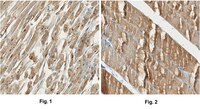
Anti-Cav1.1 alpha1s Antibody, clone 1A, Ascites Free is an antibody against Cav1.1 alpha1s for use in Western Blotting, Immunohistochemistry (Paraffin), Immunocytochemistry, Immunofluorescence, Immunoprecipitation.
Key Applications
  • Western Blotting
  • Immunohistochemistry (Paraffin)
  • Immunocytochemistry
  • Immunofluorescence
  • Immunoprecipitation
Application NotesWestern Blotting Analysis: 1.0 µg/mL of this antibody detected Cav1.1 alpha1s in 10 µg of HEK 293 and Raji membrane extracts.
Immunohistochemistry Analysis: A 1:250 dilution from a representative lot detected Cav1.1 alpha1s in human heart and rat skeletal muscle tissue sections.
Immunocytochemistry Analysis: Representative lots detected Cav1.1 localization by fluorescent immunocytochemistry staining of paraformaldehyde-fixed and Triton X-100-permeabilized mouse C2C12 myotubes as well as myotubes from primary mouse fetal skeletal myoblasts in culture (Couchoux, H., et al. (2011). Int .J. Biochem. Cell Biol. 43(5):713-720; Couchoux, H., et al. (2007). J. Physiol. 580(Pt.3):745-754).
Immunocytochemistry Analysis: A representative lot detected an upregulated Cav1.1 (α1s) surface expression on differentiating H9C2 rat ventricular myoblasts by fluorescent immunocytochemistry with or without cell permeabilization (Ménard, C., et al. (1999). J. Biol. Chem. 274(41):29063-29070).
Immunofluorescence Analysis: A representative lot immunostained T-tubule membrane in rat soleus muscle longitudinal cryosections by fluorescent immunohistochemistry (Zanin, M., et al. (2008). Am. J. Physiol. Cell Physiol. 294(1):C36-C46).
Immunohistochemistry Analysis: A representative lot immunostained the raphide and druse crystal idioblasts in paraffin-embedded sections of young Pistia stratiotes leaves (Volk, G.M., et al. (2004). Ann. Bot. 93(6):741-753).
Immunoprecipitation Analysis: A representative lot immunoprecipitated L-type Ca2+ channel Cav1.1 from mouse extensor digitorum longus muscle fiber homogenates (Delbono, O., et al. (1997). J. Neurosci. 17(18):6918-6928).
Immunoprecipitation Analysis: A representative lot immunoprecipitated dihydropyridine PN200-100-binding complex from rabbit skeletal muscle transverse tubul (T-tubule) membrane preparation with an estimated affinity of 5 nM (Morton, M. E., and Froehner, S. C. (1987). J. Biol. Chern. 262(25):11904-11907).
Function Analysis: A representative lot, when applied externally to BC3H1 mouse muscle cells, attentuated the slowly-activating, DHP-sensitive, high-threshold calcium current in a concentration-dependent manner (Morton, M.E., et al. (1988). J. Biol. Chem. 263(2):613-616).
Immunoaffinity Purification Analysis: A representative lot was conjugated to resins and used to further purify the dihydropyridine (DHP) PN200-100-binding complex isolated by wheat germ agglutinin (WGA) chromatography from rabbit skeletal muscle transverse tubul (T-tubule) membrane preparation (Morton, M. E., and Froehner, S. C. (1987). J. Biol. Chern. 262(25):11904-11907).
Western Blotting Analysis: Representative lots detected Cav1.1 alpha 1 subunit in skeletal & hart muscle tissues from rats and mice, as well as as well as in differentiating mouse C2C12 and rat H9C2 myoblasts (Esposito, A., et al. (2007). J. Appl. Physiol. 102(4):1640-1648; Bidaud, I., et al. (2006). J. Muscle Res. Cell Motil. 27(1):75-81; Ménard, C., et al. (1999). J. Biol. Chem. 274(41):29063-29070; Delbono, O., et al. (1997). J. Neurosci. 17(18):6918-6928).
Western Blotting Analysis: A representative lot detected a ~180 kDa Ca2+ channel‐like protein in the microsomal membrane preparation from Pistia stratiotes leaves (Volk, G.M., et al. (2004). Ann. Bot. 93(6):741-753).
Western Blotting Analysis: A representative lot detected the alpha 1 subunit of dihydropyridine receptor (DHPR) in rat tibialis anterior (TA) and rabbit hip adductor muscle extracts (Damiani, E., et al. (1996). Cell Calcium. 19(1):15-27).
Western Blotting Analysis: A representative lot detected a ~210 kDa target band from BC3H1 mouse muscle cell membrane extract (Morton, M.E., et al. (1988). J. Biol. Chem. 263(2):613-616).
Western Blotting Analysis: A representative lot detected a ~200 kDa target band under both reducing or non-reducing conditions using the dihydropyridine (DHP) PN200-100-binding complex isolated from rabbit skeletal muscle transverse tubul (T-tubule) membrane preparation by wheat germ agglutinin (WGA) column (Morton, M. E., and Froehner, S. C. (1987). J. Biol. Chern. 262(25):11904-11907).
Biological Information
ImmunogenDihydropyridine-binding complex isolated from rabbit muscle t-tubules.
EpitopeExtracellular domain.
Clone1A
ConcentrationPlease refer to lot specific datasheet.
HostMouse
SpecificityClone 1A targets the extracellular region of voltage-dependent L-type calcium channel subunit alpha-1S or Cav1.1 encoded by the CACNA1S gene.
IsotypeIgG1κ
Species Reactivity
  • Human
  • Mouse
  • Rabbit
  • Green Plants
Antibody TypeMonoclonal Antibody
Entrez Gene Number
Gene Symbol
  • CACNA1S
  • CACH1
  • CACN1
  • CACNL1A3
Purification MethodProtein G Purified
UniProt Number
Molecular Weight~212 kDa observed. Uncharacterized band(s) may appear in some lysates.
Physicochemical Information
Dimensions
Materials Information
Toxicological Information
Safety Information according to GHS
Safety Information
Product Usage Statements
Quality AssuranceEvaluated by Western Blotting in Jurkat membrane extract.

Western Blotting Analysis: 1.0 µg/mL of this antibody detected Cav1.1 alpha1s in 10 µg of Jurkat membrane extract.
Usage Statement
  • Unless otherwise stated in our catalog or other company documentation accompanying the product(s), our products are intended for research use only and are not to be used for any other purpose, which includes but is not limited to, unauthorized commercial uses, in vitro diagnostic uses, ex vivo or in vivo therapeutic uses or any type of consumption or application to humans or animals.
Storage and Shipping Information
Storage ConditionsStable for 1 year at -20°C from date of receipt.
Handling Recommendations: Upon receipt and prior to removing the cap, centrifuge the vial and gently mix the solution. Aliquot into microcentrifuge tubes and store at -20°C. Avoid repeated freeze/thaw cycles, which may damage IgG and affect product performance.
Packaging Information
Material Size100 μg
Transport Information
Supplemental Information
Specifications
Global Trade Item Number
Numero di catalogo GTIN
MAB427-C 04055977334258