


DR1025 Sigma-AldrichPhosphoDetect™ Anti-Chk1 (pSer³¹⁷) Rabbit pAb
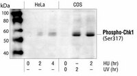

This PhosphoDetect™ Anti-Chk1 (pSer³¹⁷) Rabbit pAb is validated for use in Immunoblotting, Immunocytochemistry, Paraffin Sections for the detection of Chk1 (pSer³¹⁷).
More>> This PhosphoDetect™ Anti-Chk1 (pSer³¹⁷) Rabbit pAb is validated for use in Immunoblotting, Immunocytochemistry, Paraffin Sections for the detection of Chk1 (pSer³¹⁷). Less<<FDS (Fiches de données de sécurité), certificats d’analyse (CoA) et de qualité (CoQ), dossiers, brochures et autres documents disponibles.
Produits recommandés


Aperçu
| Replacement Information |
|---|
Tableau de caractéristiques principal
| Species Reactivity | Host | Antibody Type |
|---|---|---|
| H, Mk, M, R | Rb | Polyclonal Antibody |
Prix & Disponibilité
| Référence | Disponibilité | Conditionnement | Qté | Prix | Quantité | |
|---|---|---|---|---|---|---|
| DR1025-50UL |
|
Ampoule plast. | 50 ul |
|
— |

| Product Information | |
|---|---|
| Form | Liquid |
| Formulation | In 150 mM NaCl, 10 mM HEPES, 100 µg/ml BSA, 50% glycerol, pH 7.5. |
| Positive control | Lysates from COS cells treated with UV |
| Preservative | None |
| Quality Level | MQ100 |
| Physicochemical Information |
|---|
| Dimensions |
|---|
| Materials Information |
|---|
| Toxicological Information |
|---|
| Safety Information according to GHS |
|---|
| Safety Information |
|---|
| Product Usage Statements |
|---|
| Storage and Shipping Information | |
|---|---|
| Ship Code | Blue Ice Only |
| Toxicity | Standard Handling |
| Storage | -20°C |
| Do not freeze | Ok to freeze |
| Special Instructions | Do not aliquot. |
| Packaging Information |
|---|
| Transport Information |
|---|
| Supplemental Information |
|---|
| Specifications |
|---|
| Global Trade Item Number | |
|---|---|
| Référence | GTIN |
| DR1025-50UL | 04055977225914 |
Documentation
PhosphoDetect™ Anti-Chk1 (pSer³¹⁷) Rabbit pAb FDS
| Titre |
|---|
PhosphoDetect™ Anti-Chk1 (pSer³¹⁷) Rabbit pAb Certificats d'analyse
| Titre | Numéro de lot |
|---|---|
| DR1025 |
Références bibliographiques
| Aperçu de la référence bibliographique |
|---|
| Zhao, H. and Piwnica-Worms, H. 2001. Mol. Cell. Biol. 21, 4129. Shieh, S.Y., et al. 2000. Genes Dev. 14, 289. Martinho, R.G., et al. 1998. EMBO J. 17, 7239. Zeng, Y., et al. 1998. Nature 395, 507. |