Millipore Sigma Vibrant Logo

MAB1393-I Anti-PECAM-1 (CD31) Antibody, clone TLD-3A12

View Products on Sigmaaldrich.com
MAB1393-I
100 μg  
Prix en cours de récupération
Le prix n'a pas pu être récupéré
La quantité minimale doit être un multiple de
Maximum Quantity is
À la validation de la commande Plus d'informations
Vous avez sauvegardé ()
 
Demander le prix
Disponibilité limitée
En stock 
Interrompu(e)
Disponible en quantités limitées
Disponibilité à confirmer
    Pour le restant : Nous vous tiendrons informé
      Pour le restant : Nous vous tiendrons informé
      Nous vous tiendrons informé
      Contacter le Service Clients
      Contact Customer Service


      Contacter le Service Clients

      Aperçu

      Replacement Information

      Tableau de caractéristiques principal

      Species ReactivityKey ApplicationsHostFormatAntibody Type
      H, RIHC, FUNC, WB, FC, ELISA, IH(P)MPurifiedMonoclonal Antibody
      Description
      Catalogue NumberMAB1393-I
      DescriptionAnti-PECAM-1 (CD31) Antibody, clone TLD-3A12
      Alternate Names
      • Platelet endothelial cell adhesion molecule
      • CD31
      Background InformationPlatelet endothelial cell adhesion molecule (UniProt: Q3SWT0; PECAM-1, CD31) is encoded by the Pecam1 (also known as Pecam) gene (Gene ID: 29583) in rat.PECAM-1 is a single-pass type I membrane protein that contains 6 Ig-like C2-type (immunoglobulin-like) domains. PECAM1 is differentially glycosylated involving both N-linked and O-linked glycosylation sites. It is a cell adhesion molecule that is required for leukocyte trans-endothelial migration (TEM) under most inflammatory conditions. PECAM1 plays a vital role in signaling processes involved in angiogenesis, platelet function, thrombosis, mechanosensing of endothelial cell response to fluid shear stress, and regulation of multiple stages of leukocyte migration through venular walls. PECAM1 is expressed on platelets and leukocytes and is primarily concentrated at the borders between endothelial cells. PECAM1 prevents phagocyte ingestion of closely apposed viable cells by transmitting 'detachment' signals, and changes function on apoptosis, promoting tethering of dying cells to phagocytes. During apoptosis, the inside-out signaling of PECAM1 is disabled that allows the apoptotic cell to accept phagocyte interaction. PECAM1 is phosphorylated on serine and tyrosine residues following cellular activation. In endothelial cells Fyn is shown to mediate mechanical-force induced tyrosine phosphorylation. In response to Fc epsilon RI (FCER1 activation), PECAM1 is phosphorylated on tyrosine residues by FER and FES.
      References
      Product Information
      FormatPurified
      PresentationPurified mouse monoclonal antibody IgG1 in PBS without azide.
      Quality LevelMQ100
      Applications
      Application
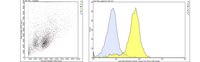
This mouse monoclonal Anti-PECAM-1 (CD31), clone TLD-3A12, Cat. No. MAB1393-I is tested for use in ELISA, Flow Cytometry, Functional Studies, Immunohistochemistry (Paraffin) and Immunohistochemistry, Immunoprecipitation, and Western Blotting, for the detection of CD31/PECAM-1.
      Key Applications
      • Immunohistochemistry
      • Affects Function
      • Western Blotting
      • Flow Cytometry
      • ELISA
      • Immunohistochemistry (Paraffin)
      Application NotesImmunoprecipitation Analysis: A representative lot detected PECAM-1 (CD31) in Immunoprecipitation applications (Williams, K.C., et. al. (1996). J Neurosci Res. 45(6):747-57).

      Flow Cytometry Analysis: 1 µg from a representative lot detected PECAM-1 (CD31) in one million rat splenocytes.

      Western Blotting Analysis: A representative lot detected PECAM-1 (CD31) in Western Blotting applications (Male, D., et. al. (1995). Immunology. 84(3):453-60).

      ELISA Analysis: A representative lot detected PECAM-1 (CD31) in ELISA applications (Male, D., et. al. (1995). Immunology. 84(3):453-60; Williams, K.C., et. al. (1996). J Neurosci Res. 45(6):747-57).

      Affects Function: A representative lot of PECAM-1 (CD31) Affected Function (Williams, K.C., et. al. (1996). J Neurosci Res. 45(6):747-57).

      Immunohistochemistry Analysis: A representative lot detected PECAM-1 (CD31) in Immunohistochemistry applications (Williams, K.C., et. al. (1996). J Neurosci Res. 45(6):747-57).
      Biological Information
      ImmunogenLewis rat derived activated microglial cells.
      CloneTLD-3A12
      ConcentrationPlease refer to lot specific datasheet.
      HostMouse
      SpecificityClone TLD-3A12 recognizes rat and human PECAM-1.
      IsotypeIgG1κ
      Species Reactivity
      • Human
      • Rat
      Species Reactivity NoteHuman, Rat.
      Antibody TypeMonoclonal Antibody
      Entrez Gene Number
      Gene Symbol
      • Pecam1
      • Pecam
      Purification MethodProtein G purified
      UniProt Number
      Molecular Weight76.19 kDa calculated.
      Physicochemical Information
      Dimensions
      Materials Information
      Toxicological Information
      Safety Information according to GHS
      Safety Information
      Product Usage Statements
      Quality AssuranceEvaluated by Immunohistochemistry in human tonsil.

      Immunohistochemistry Analysis: A 1:250 dilution of this antibody detected PECAM-1 (CD31) in human tonsil tissue.
      Usage Statement
      • Unless otherwise stated in our catalog or other company documentation accompanying the product(s), our products are intended for research use only and are not to be used for any other purpose, which includes but is not limited to, unauthorized commercial uses, in vitro diagnostic uses, ex vivo or in vivo therapeutic uses or any type of consumption or application to humans or animals.
      Storage and Shipping Information
      Storage ConditionsStable for 1 year at -20°C from date of receipt. Handling Recommendations: Upon receipt and prior to removing the cap, centrifuge the vial and gently mix the solution. Aliquot into microcentrifuge tubes and store at -20°C. Avoid repeated freeze/thaw cycles, which may damage IgG and affect product performance.
      Packaging Information
      Material Size100 μg
      Transport Information
      Supplemental Information
      Specifications
      Global Trade Item Number
      Référence GTIN
      MAB1393-I 04054839199325

      Documentation

      Anti-PECAM-1 (CD31) Antibody, clone TLD-3A12 FDS

      Titre

      Fiche de données de sécurité des matériaux (FDS) 

      Anti-PECAM-1 (CD31) Antibody, clone TLD-3A12 Certificats d'analyse

      TitreNuméro de lot
      Anti-PECAM-1 (CD31), clone TLD-3A12 - 3308162 3308162
      Anti-PECAM-1 (CD31), clone TLD-3A12 - 3526285 3526285
      Anti-PECAM-1 (CD31), clone TLD-3A12 - 4045766 4045766
      Anti-PECAM-1 (CD31), clone TLD-3A12 - 4109622 4109622
      Anti-PECAM-1 (CD31), clone TLD-3A12 Monoclonal Antibody 3036819
      Anti-PECAM-1 (CD31), clone TLD-3A12 Monoclonal Antibody 3096939
      Anti-PECAM-1 (CD31), clone TLD-3A12, Monoclonal Antibody Q2841548