Millipore Sigma Vibrant Logo

GR36 Anti-Insulin Receptor (β-Subunit) Mouse mAb (CT-3)

View Products on Sigmaaldrich.com
GR36
  
Prix en cours de récupération
Le prix n'a pas pu être récupéré
La quantité minimale doit être un multiple de
Maximum Quantity is
À la validation de la commande Plus d'informations
Vous avez sauvegardé ()
 
Demander le prix
Disponibilité limitée
En stock 
Interrompu(e)
Disponible en quantités limitées
Disponibilité à confirmer
    Pour le restant : Nous vous tiendrons informé
      Pour le restant : Nous vous tiendrons informé
      Nous vous tiendrons informé
      Contacter le Service Clients
      Contact Customer Service


      Contacter le Service Clients

      Aperçu

      Replacement Information

      Tableau de caractéristiques principal

      Species ReactivityHostAntibody Type
      H, M, RMMonoclonal Antibody
      Description
      OverviewRecognizes the ~95 kDa the β-subunit of insulin receptor in IM9 cells, placenta, and fetal liver tissue.
      Catalogue NumberGR36
      Brand Family Calbiochem®
      Application Data
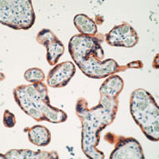
      Detection of human insulin receptor (β-subunit) by immunohistochemistry. Sample: Human placenta tissue fixed in formalin and embedded in paraffin. Primary antibody: Anti-Insulin Receptor (β-Subunit) Mouse mAb (CT-3) (Cat. No. GR36) (1:50). Detection: fluorescence.

      Detection of human insulin receptor (β-subunit) by immunoblotting. Sample: Human insulin receptor. Primary antibody: Anti-Insulin Receptor (β-Subunit) Mouse mAb (CT-3) (Cat. No. GR36) (1 µg/ml). Detection: chemiluminescence.
      References
      ReferencesGrigorescu, F., et al. 1987. J.Clin. Endocrinol. Metab. 64, 549.
      Hari, J. and Roth, R.A. 1987. J. Biol. Chem. 262, 15341.
      Rosen, O.M. 1987. Science 237, 1452.
      Morgan, D.O., et al. 1986. Proc. Natl. Acad. Sci. USA 83, 328.
      Morgan, D.O. and Roth, R.A. 1986. Biochemistry 25, 1364.
      White, M.F. and Kahn, C.R. 1986. The Enzymes 17, 247.
      Ganguly, S., et al. 1985. In Current Topics in Cellular Regulation, Academic Press 27, 83.
      Grunberger, G., et al. 1984. Science 223, 932.
      Product Information
      FormLiquid
      FormulationIn 10 mM PBS, 0.2% BSA, pH 7.4.
      Positive controlIM-9 lymphocyte cells or placenta or liver tissue
      Preservative≤0.1% sodium azide
      Quality LevelMQ100
      Applications
      Key Applications Frozen Sections
      Immunoblotting (Western Blotting)
      Not Immunoprecipitation
      Paraffin Sections
      Application NotesFrozen Sections (1:50-1:100)
      Immunoblotting (1:200-1:400)
      Immunoprecipitation (not recommended)
      Paraffin Sections (1:50-1:100, pressure cooker pre-treatment required)
      Application CommentsNo cross-reactivity with IGF receptors. Will stain IM-9 lymphocyte cells as well as placenta and liver tissue. This antibody is also useful for tyrosine kinase assays for antibody mediated capture on microwell plates. Antibody should be titrated for optimal results in individual systems.
      Biological Information
      Immunogena recombinant protein consisting of the C-terminal 100 amino acids of the β-subunit of human insulin receptor fused to GST
      ImmunogenHuman
      CloneCT-3
      HostMouse
      IsotypeIgG₁
      Species Reactivity
      • Human
      • Mouse
      • Rat
      Antibody TypeMonoclonal Antibody
      Concentration Label Please refer to vial label for lot-specific concentration
      Physicochemical Information
      Dimensions
      Materials Information
      Toxicological Information
      Safety Information according to GHS
      Safety Information
      Product Usage Statements
      Storage and Shipping Information
      Ship Code Blue Ice Only
      Toxicity Standard Handling
      Storage +2°C to +8°C
      Avoid freeze/thaw Avoid freeze/thaw
      Do not freeze Ok to freeze
      Special InstructionsFollowing initial use, aliquot and freeze (-20°C) for long-term storage. Avoid freeze/thaw cycles. Centrifuge the contents of the vial containing the antibody before use to remove any solution that may have accumulated around or under the cap of the vial during shipment or storage.
      Packaging Information
      Transport Information
      Supplemental Information
      Specifications
      Global Trade Item Number
      Référence GTIN
      GR36 0

      Documentation

      Anti-Insulin Receptor (β-Subunit) Mouse mAb (CT-3) FDS

      Titre

      Fiche de données de sécurité des matériaux (FDS) 

      Anti-Insulin Receptor (β-Subunit) Mouse mAb (CT-3) Certificats d'analyse

      TitreNuméro de lot
      GR36

      Références bibliographiques

      Aperçu de la référence bibliographique
      Grigorescu, F., et al. 1987. J.Clin. Endocrinol. Metab. 64, 549.
      Hari, J. and Roth, R.A. 1987. J. Biol. Chem. 262, 15341.
      Rosen, O.M. 1987. Science 237, 1452.
      Morgan, D.O., et al. 1986. Proc. Natl. Acad. Sci. USA 83, 328.
      Morgan, D.O. and Roth, R.A. 1986. Biochemistry 25, 1364.
      White, M.F. and Kahn, C.R. 1986. The Enzymes 17, 247.
      Ganguly, S., et al. 1985. In Current Topics in Cellular Regulation, Academic Press 27, 83.
      Grunberger, G., et al. 1984. Science 223, 932.
      Fiche technique

      Note that this data sheet is not lot-specific and is representative of the current specifications for this product. Please consult the vial label and the certificate of analysis for information on specific lots. Also note that shipping conditions may differ from storage conditions.

      Revision27-August-2007 RFH
      ApplicationFrozen Sections (1:50-1:100)
      Immunoblotting (1:200-1:400)
      Immunoprecipitation (not recommended)
      Paraffin Sections (1:50-1:100, pressure cooker pre-treatment required)
      Application Data
      Detection of human insulin receptor (β-subunit) by immunohistochemistry. Sample: Human placenta tissue fixed in formalin and embedded in paraffin. Primary antibody: Anti-Insulin Receptor (β-Subunit) Mouse mAb (CT-3) (Cat. No. GR36) (1:50). Detection: fluorescence.

      Detection of human insulin receptor (β-subunit) by immunoblotting. Sample: Human insulin receptor. Primary antibody: Anti-Insulin Receptor (β-Subunit) Mouse mAb (CT-3) (Cat. No. GR36) (1 µg/ml). Detection: chemiluminescence.
      DescriptionPurified mouse monoclonal antibody gernerated by immunizing BALB/c mice with the specified immunogen and fusing splenocytes with NS1 mouse myeloma cells. Recognizes the ~95 kDa the β-subunit of insulin receptor.
      BackgroundInsulin initiates its effects on cells through interaction with high-affinity, cell surface, glycoprotein receptors, consisting of two α (~135 kDa) and two β (~95 kDa) subunits linked by disulfide bonds. The α subunits, which are completely extracellular, bind insulin and the β subunits possessing a single transmembrane domain become phosphorylated in response to insulin. Insulin-dependent phosphorylation occurs at multiple sites near the carboxyl terminus and in the kinase domain. The receptor protein-tyrosine kinase has been implicated as the mediator of most, if not all, of the effects of insulin. Mutations that diminish both phosphorylation and internalization of the insulin receptor block insulin action, as do antibodies, which inhibit its protein-tyrosine kinase activity. Cells from patients with diabetic syndromes, characterized by non-responsiveness to insulin, exhibit reduced insulin dependent protein-tyrosine kinase activity.
      HostMouse
      Immunogen speciesHuman
      Immunogena recombinant protein consisting of the C-terminal 100 amino acids of the β-subunit of human insulin receptor fused to GST
      CloneCT-3
      IsotypeIgG₁
      Specieshuman, mouse, rat
      Positive controlIM-9 lymphocyte cells or placenta or liver tissue
      FormLiquid
      FormulationIn 10 mM PBS, 0.2% BSA, pH 7.4.
      Concentration Label Please refer to vial label for lot-specific concentration
      Preservative≤0.1% sodium azide
      CommentsNo cross-reactivity with IGF receptors. Will stain IM-9 lymphocyte cells as well as placenta and liver tissue. This antibody is also useful for tyrosine kinase assays for antibody mediated capture on microwell plates. Antibody should be titrated for optimal results in individual systems.
      Storage Avoid freeze/thaw
      +2°C to +8°C
      Do Not Freeze Ok to freeze
      Special InstructionsFollowing initial use, aliquot and freeze (-20°C) for long-term storage. Avoid freeze/thaw cycles. Centrifuge the contents of the vial containing the antibody before use to remove any solution that may have accumulated around or under the cap of the vial during shipment or storage.
      Toxicity Standard Handling
      ReferencesGrigorescu, F., et al. 1987. J.Clin. Endocrinol. Metab. 64, 549.
      Hari, J. and Roth, R.A. 1987. J. Biol. Chem. 262, 15341.
      Rosen, O.M. 1987. Science 237, 1452.
      Morgan, D.O., et al. 1986. Proc. Natl. Acad. Sci. USA 83, 328.
      Morgan, D.O. and Roth, R.A. 1986. Biochemistry 25, 1364.
      White, M.F. and Kahn, C.R. 1986. The Enzymes 17, 247.
      Ganguly, S., et al. 1985. In Current Topics in Cellular Regulation, Academic Press 27, 83.
      Grunberger, G., et al. 1984. Science 223, 932.