Le fait de fermer ne sauvegardera pas votre configuration à moins que vous n'ajoutiez l'article à votre Panier d'achat ou à vos Favoris.
Cliquer sur OK pour fermer l'outil MILLIPLEX® MAP ou sur Annuler pour retourner à votre sélection.
Choisissez des Panels configurables & des Kits préconfigurés - OU - des MAPmate™ de signalisation cellulaire
Concevez vos kits MILLIPLEX® MAP et obtenez leur prix.
Panels configurables & Kits préconfigurés
Notre large gamme est constituée de panels multiplex qui vous permettent de choisir, au sein d'un panel, les analytes qui répondent le mieux à vos besoins. Sur un autre onglet, vous pouvez choisir un format cytokine préconfiguré ou un kit Simplex.
Kits de signalisation cellulaire & MAPmate™
Choisissez des kits préconfigurés qui permettent d'explorer l'ensemble des voies ou des processus. Ou concevez vos propres kits en choisissant des Simplex MAPmate™ et en suivant les instructions fournies.
Les MAPmate™ suivants ne peuvent pas être utilisés ensemble : -des MAPmate™ qui nécessitent des tampons différents -des paires de MAPmate™ totaux et phospho-spécifiques, par ex. GSK3β total et GSK3β (Ser 9) -des MAPmate™ PanTyr et spécifiques d'un site, par ex. Récepteur Phospho-EGF et phospho-STAT1 (Tyr701) -Plus d'un phospho-MAPmate™ pour une seule cible (Akt, STAT3). -GAPDH et β-Tubuline ne peuvent pas être utilisés avec les kits ou les MAPmate™ contenant panTyr.
.
Référence
Guide d'achat
Qté
Liste
Cet article a été ajouté à vos favoris.
Sélectionner une espèce, un type de panel, un kit ou un type d'échantillon
Pour commencer à concevoir votre kit MILLIPLEX® MAP, sélectionnez une espèce, un type de panel ou un kit d'intérêt.
Custom Premix Selecting "Custom Premix" option means that all of the beads you have chosen will be premixed in manufacturing before the kit is sent to you.
Catalogue Number
Ordering Description
Qty/Pack
List
Cet article a été ajouté à vos favoris.
Espèce
Type de panel
Kit sélectionné
Qté
Référence
Guide d'achat
Qté
Prix tarif
96-Well Plate
Qté
Référence
Guide d'achat
Qté
Prix tarif
Ajouter des réactifs supplémentaires (Un kit "Buffer and Detection Kit" est nécessaire pour une utilisation avec les MAPmate™)
Qté
Référence
Guide d'achat
Qté
Prix tarif
48-602MAG
Buffer Detection Kit for Magnetic Beads
1 Kit
Option de gain de place Nos clients qui commandent plusieurs kits peuvent choisir d'économiser de l'espace de stockage en éliminant l'emballage de chaque kit et de recevoir les composants de leur essai multiplex conditionnés sous poches en plastique pour un stockage plus compact.
Cet article a été ajouté à vos favoris.
Ce produit a été ajouté à votre panier.
Vous pouvez maintenant concevoir un autre kit personnalisé, choisir un kit pré-configuré, régler vos achats ou fermer l'outil de commande.
NE1015
Sigma-AldrichAnti-Glial Fibrillary Acidic Protein Cocktail Mouse mAb (SMI-22)
This Anti-Glial Fibrillary Acidic Protein Cocktail Mouse mAb is validated for use in ELISA, Frozen Sections, WB, ICC, Paraffin Sections for the detection of Glial Fibrillary Acidic Protein.
More>>This Anti-Glial Fibrillary Acidic Protein Cocktail Mouse mAb is validated for use in ELISA, Frozen Sections, WB, ICC, Paraffin Sections for the detection of Glial Fibrillary Acidic Protein. Less<<
FDS (Fiches de données de sécurité), certificats d’analyse (CoA) et de qualité (CoQ), dossiers, brochures et autres documents disponibles.
Le champ quantité est vide. Veuillez saisir une quantité supérieure ou égale à 1 au minimum pour ajouter des articles à votre panier.
Description
Overview
Recognizes ~50 kDa glial fibrillary acidic protein (GFAP) in human and bovine cytoskeletal preparations.
Catalogue Number
NE1015
Brand Family
Calbiochem®
Synonyms
Anti-GFAP Cocktail
Application Data
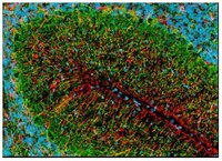
Detection of rat glial fibrillary acidic protein by staining frozen sections. Sample: Rat brain. Primary antibody: Anti-GFAP Cocktail Mouse mAb (SMI-22) (Cat. No. NE1015) (1:1000). Detection: fluorescence (red) with Hoechst 33342 counterstain. Primary mixed glial cultures were stained with anti-mouse GFAP (EMD Millipore, Cat. No. MAB360) and a DyLight™488 conjugated goat anti-mouse secondary Antibody. The nucleus was counterstained with DAPI. The image was captured using Olympus BX51 with an exposure time of 982 millisec for green channel and 126 millisec for DAPI channel. Magnification is 10X without any correction on Gain/Offset/Bad pixel.
Courtesy of Jose Shinsmon, Immunology, Dept of Pathology, Faculty of Medicine and Health Sciences, UPM,Serdang 43400.
References
References
Vick, W.W., et al. 1987. Acta. Cytol.31, 816. McLendon R.E., et al. 1986. J. Neuropathol. Exp. Neurol.45, 692. Pegram, C.N., et al. 1985. Neurochem. Pathol.3, 119.
ELISA (1:1000) Frozen Sections (1:1000, see comments) Immunoblotting (1:1000) Immunocytochemistry (1:1000, see comments) Paraffin Sections (1:1000, trypsin or heat pre-treatment required)
Application Comments
This cocktail is derived from the Bigner-Eng clones MAb1B4, MAb2E1, and MAb4A11 and provides a means for more comprehensive detection of astrocytomas than each clone alone. Each component is specific for GFAP and stains astrocytes and astrocytic processes as well as Bergman glia. Recognizes both anaplastic and reactive astrocytes by immunocytochemical staining. Does not recognize metastatic tumors and brain tumors of non-astrocytic origin, including medulloblastomas, meningiomas, choroid plexus papillomas, and schwannomas. For staining paraffin sections it is recommended that de-paraffinized sections be treated with 0.1% trypsin in 50 mM Tris-HCl, pH 7.6 for 20-30 min at 37°C or boiled in Tris-buffered saline, pH 9.0 for 15 min to expose the epitope. For immunocytochemistry or staining frozen sections, post-fixation in cold methanol or methanol/hydrogen peroxide for 10 min is required for access to the astrocytes in the sample. Antibody should be titrated for optimal results in individual systems.
Biological Information
Immunogen
purified bovine GFAP protein
Immunogen
Bovine
Clone
SMI-22
Host
Mouse
Isotype
IgG2b
Species Reactivity
Bovine
Canine
Chicken
Guinea Pig
Human
Mouse
Porcine
Rat
Sheep
Antibody Type
Monoclonal Antibody
Storage and Shipping Information
Ship Code
Dry Ice Only
Toxicity
Standard Handling
Storage
-20°C
Avoid freeze/thaw
Avoid freeze/thaw
Do not freeze
Ok to freeze
Special Instructions
Upon initial thaw, aliquot and freeze (-20°C).
Global Trade Item Number
Référence
GTIN
NE1015-100UL
04055977209853
Documentation
Anti-Glial Fibrillary Acidic Protein Cocktail Mouse mAb (SMI-22) FDS
Anti-Glial Fibrillary Acidic Protein Cocktail Mouse mAb (SMI-22) Certificats d'analyse
Titre
Numéro de lot
NE1015
Références bibliographiques
Aperçu de la référence bibliographique
Vick, W.W., et al. 1987. Acta. Cytol.31, 816. McLendon R.E., et al. 1986. J. Neuropathol. Exp. Neurol.45, 692. Pegram, C.N., et al. 1985. Neurochem. Pathol.3, 119.
Fiche technique
Note that this data sheet is not lot-specific and is representative of the current specifications for this product. Please consult the vial label and the certificate of analysis for information on specific lots. Also note that shipping conditions may differ from storage conditions.
Revision
01-October-2007 RFH
Synonyms
Anti-GFAP Cocktail
Application
ELISA (1:1000) Frozen Sections (1:1000, see comments) Immunoblotting (1:1000) Immunocytochemistry (1:1000, see comments) Paraffin Sections (1:1000, trypsin or heat pre-treatment required)
Application Data
Detection of rat glial fibrillary acidic protein by staining frozen sections. Sample: Rat brain. Primary antibody: Anti-GFAP Cocktail Mouse mAb (SMI-22) (Cat. No. NE1015) (1:1000). Detection: fluorescence (red) with Hoechst 33342 counterstain. Primary mixed glial cultures were stained with anti-mouse GFAP (EMD Millipore, Cat. No. MAB360) and a DyLight™488 conjugated goat anti-mouse secondary Antibody. The nucleus was counterstained with DAPI. The image was captured using Olympus BX51 with an exposure time of 982 millisec for green channel and 126 millisec for DAPI channel. Magnification is 10X without any correction on Gain/Offset/Bad pixel.
Courtesy of Jose Shinsmon, Immunology, Dept of Pathology, Faculty of Medicine and Health Sciences, UPM,Serdang 43400.
Description
Mouse monoclonal antibody cocktail that contains a mixture of 3 antibodies supplied as undiluted ascites. Recognizes the ~50 kDa glial fibrillary acidic protein.
Background
Glial fibrillary acidic protein (GFAP) is an intermediate filament protein found only in glial cells or cells of glial origin. It can be detected in astrocytes and certain other astroglia in the CNS, in satellite cells in peripheral ganglia, and in non-myelinating Schwann cells in peripheral nerves. GFAP is upregulated and expressed at high levels in astrocytes in many damage and disease states. It is also often highly expressed in neural stem cells and many types of brain tumors.
Host
Mouse
Immunogen species
Bovine
Immunogen
purified bovine GFAP protein
Clone
SMI-22
Isotype
IgG2b
Species
bovine, canine, chicken, guinea pig, human, mouse, porcine, rat, sheep
Positive control
Astrocytes or cytoskeletal preparations
Form
Liquid
Formulation
Undiluted ascites.
Preservative
≤ 0.1% sodium azide
Comments
This cocktail is derived from the Bigner-Eng clones MAb1B4, MAb2E1, and MAb4A11 and provides a means for more comprehensive detection of astrocytomas than each clone alone. Each component is specific for GFAP and stains astrocytes and astrocytic processes as well as Bergman glia. Recognizes both anaplastic and reactive astrocytes by immunocytochemical staining. Does not recognize metastatic tumors and brain tumors of non-astrocytic origin, including medulloblastomas, meningiomas, choroid plexus papillomas, and schwannomas. For staining paraffin sections it is recommended that de-paraffinized sections be treated with 0.1% trypsin in 50 mM Tris-HCl, pH 7.6 for 20-30 min at 37°C or boiled in Tris-buffered saline, pH 9.0 for 15 min to expose the epitope. For immunocytochemistry or staining frozen sections, post-fixation in cold methanol or methanol/hydrogen peroxide for 10 min is required for access to the astrocytes in the sample. Antibody should be titrated for optimal results in individual systems.
Storage
Avoid freeze/thaw
-20°C
Do Not Freeze
Ok to freeze
Special Instructions
Upon initial thaw, aliquot and freeze (-20°C).
Toxicity
Standard Handling
References
Vick, W.W., et al. 1987. Acta. Cytol.31, 816. McLendon R.E., et al. 1986. J. Neuropathol. Exp. Neurol.45, 692. Pegram, C.N., et al. 1985. Neurochem. Pathol.3, 119.