


OP16L Sigma-AldrichAnti-c-ErbB2/c-Neu (Ab-4) Mouse mAb (7.16.4)


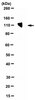
Anti-c-ErbB2/c-Neu (Ab-4), mouse monoclonal, clone 7.16.4, recognizes the ~185 kDa c-ErbB2/c-Neu in overexpressing B104-1-1 cells. It is validated for WB, ICC, IP, and IHC with frozen sections.
More>> Anti-c-ErbB2/c-Neu (Ab-4), mouse monoclonal, clone 7.16.4, recognizes the ~185 kDa c-ErbB2/c-Neu in overexpressing B104-1-1 cells. It is validated for WB, ICC, IP, and IHC with frozen sections. Less<<Anti-c-ErbB2/c-Neu (Ab-4) Mouse mAb (7.16.4): Ficha de datos de seguridad (MSDS o SDS), certificado de análisis y de calidad (CoA y CoQ), expedientes, folletos y otros documentos disponibles.
Sinónimos: Anti-HER2, Anti-Neu, Anti-ErbB2, Anti-Erythroblastosis Virus
Productos recomendados


Descripción
| Replacement Information |
|---|
Tabla espec. clave
| Species Reactivity | Host | Antibody Type |
|---|---|---|
| H, R | M | Monoclonal Antibody |
Precios y disponibilidad
| Número de referencia | Disponiblidad | Embalaje | Cant./Env. | Precio | Cantidad | |
|---|---|---|---|---|---|---|
| OP16L-100UG |
|
Frasco de vidrio | 100 μg |
|
— | |
| OP16L-500UG |
|
Frasco de vidrio | 500 μg |
|
— |
| Product Information | |
|---|---|
| Form | Lyophilized |
| Formulation | Lyophilized from a volatile buffer, 100 µg BSA. |
| Negative control | Fisher rat embryo cells |
| Positive control | B104-1-1 cells |
| Preservative | None |
| Quality Level | MQ100 |
| Physicochemical Information |
|---|
| Dimensions |
|---|
| Materials Information |
|---|
| Toxicological Information |
|---|
| Safety Information according to GHS |
|---|
| Safety Information |
|---|
| Product Usage Statements |
|---|
| Packaging Information |
|---|
| Transport Information |
|---|
| Supplemental Information |
|---|
| Specifications |
|---|
| Global Trade Item Number | |
|---|---|
| Número de referencia | GTIN |
| OP16L-100UG | 04055977208733 |
| OP16L-500UG | 04055977208740 |
Documentation
Anti-c-ErbB2/c-Neu (Ab-4) Mouse mAb (7.16.4) Certificados de análisis
| Cargo | Número de lote |
|---|---|
| OP16L |
Referencias bibliográficas
| Visión general referencias |
|---|
| Zhang, H., et al. 1999. Exp. Mol. Pathol. 67, 15. Peterson, N.C. and Greene, M.I., 1988. DNA Cell Biol. 17, 1031. Kokai, Y., et al. 1988. Proc. Natl. Acad. Sci. USA 85, 5389. Van De Vijver, M.J., et al. 1988. New Engl. J. Med. 319, 1239. DiFiore, P.P., et al. 1987. Science 237, 178. Kokai, Y., et al. 1987. Proc. Natl. Acad. Sci. USA 84, 8498. Slamon, D.J., et al. Science 235, 177. Varley, J.M., et al. 1987. Oncogene 1, 423. Bargmann, C.I., et al. 1986. Nature 319, 226. Yamamoto, T., et al. 1986. Nature 319, 230. Blick, M., et al. 1984. Blood 64, 1234. Schwab, M., et al. 1984. Cold Spring Harbor Laboratory 2, 215. |
Citas
| Título | |
|---|---|
|
|
| Ficha técnica | ||||||||||||||||||||||||||||||||||||||||||||||||||
|---|---|---|---|---|---|---|---|---|---|---|---|---|---|---|---|---|---|---|---|---|---|---|---|---|---|---|---|---|---|---|---|---|---|---|---|---|---|---|---|---|---|---|---|---|---|---|---|---|---|---|
|
Note that this data sheet is not lot-specific and is representative of the current specifications for this product. Please consult the vial label and the certificate of analysis for information on specific lots. Also note that shipping conditions may differ from storage conditions.
|