Derivation of transgene-free human induced pluripotent stem cells from human peripheral T cells in defined culture conditions.
Kishino, Y; Seki, T; Fujita, J; Yuasa, S; Tohyama, S; Kunitomi, A; Tabei, R; Nakajima, K; Okada, M; Hirano, A; Kanazawa, H; Fukuda, K
PloS one
9
e97397
2014
Mostrar resumen
Recently, induced pluripotent stem cells (iPSCs) were established as promising cell sources for revolutionary regenerative therapies. The initial culture system used for iPSC generation needed fetal calf serum in the culture medium and mouse embryonic fibroblast as a feeder layer, both of which could possibly transfer unknown exogenous antigens and pathogens into the iPSC population. Therefore, the development of culture systems designed to minimize such potential risks has become increasingly vital for future applications of iPSCs for clinical use. On another front, although donor cell types for generating iPSCs are wide-ranging, T cells have attracted attention as unique cell sources for iPSCs generation because T cell-derived iPSCs (TiPSCs) have a unique monoclonal T cell receptor genomic rearrangement that enables their differentiation into antigen-specific T cells, which can be applied to novel immunotherapies. In the present study, we generated transgene-free human TiPSCs using a combination of activated human T cells and Sendai virus under defined culture conditions. These TiPSCs expressed pluripotent markers by quantitative PCR and immunostaining, had a normal karyotype, and were capable of differentiating into cells from all three germ layers. This method of TiPSCs generation is more suitable for the therapeutic application of iPSC technology because it lowers the risks associated with the presence of undefined, animal-derived feeder cells and serum. Therefore this work will lead to establishment of safer iPSCs and extended clinical application. | 24824994
 |
SSEA-3 as a novel amplifying cancer cell surface marker in colorectal cancers.
Suzuki, Y; Haraguchi, N; Takahashi, H; Uemura, M; Nishimura, J; Hata, T; Takemasa, I; Mizushima, T; Ishii, H; Doki, Y; Mori, M; Yamamoto, H
International journal of oncology
42
161-7
2013
Mostrar resumen
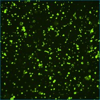
Findings from studies on stem cells have been applied to cancer stem cell (CSC) research, but little is known about the relationship between ES cell-related cell surface markers and CSCs. In this study, we focused on stage-specific embryonic antigen 3 (SSEA-3), a marker of mesenchymal stem cells and Muse cells in colorectal cancer (CRC). Expression of SSEA-3 in human CRC cell lines and clinical specimens, specifically the relationship of SSEA-3 expression and the representative CSC markers (CD44, CD166, ALDH, CD24 and CD26) as well as with mesenchymal stem cell/Muse cell marker (CD105) were assessed. To characterize SSEA-3-expressing cells, tumorigenicity, sphere formation ability, expression of iPS genes (Oct4, NANOG, SOX2 and c-Myc), cell proliferation and cell cycle status were assessed. SSEA-3 expression was identified in Caco-2, DLD-1, HT-29, SW480 and HCT116, but not in CaR-1 cells. No significant relationship between SSEA-3 and other stem cell markers was detected. SSEA-3+ cells showed increased tumorigenicity in vivo, but lower sphere formation ability in vitro than SSEA-3-. iPS gene expression was not correlated with SSEA-3 expression status. SSEA-3+ cells showed higher proliferative ability than SSEA-3- through enhanced cell cycles by decreased expression of p21Cip1/Waf1 and p27Kip1. Immunofluorescence analysis in clinical specimens indicated that expression of SSEA-3 is limited to stromal cells in normal mucosa but broad in poorly differentiated adenocarcinoma. These observations indicated that SSEA-3+ cells in CRC have immature phenotype but decreased self-renewal ability and may function as tumor transient amplifying cells or delayed contributing tumor-initiating cells. | 23175153
|
Induction of pluripotent stem cells from human third molar mesenchymal stromal cells.
Oda, Y; Yoshimura, Y; Ohnishi, H; Tadokoro, M; Katsube, Y; Sasao, M; Kubo, Y; Hattori, K; Saito, S; Horimoto, K; Yuba, S; Ohgushi, H
The Journal of biological chemistry
285
29270-8
2009
Mostrar resumen
The expression of four transcription factors (OCT3/4, SOX2, KLF4, and MYC) can reprogram mouse as well as human somatic cells to induced pluripotent stem (iPS) cells. We generated iPS cells from mesenchymal stromal cells (MSCs) derived from human third molars (wisdom teeth) by retroviral transduction of OCT3/4, SOX2, and KLF4 without MYC, which is considered as oncogene. Interestingly, some of the clonally expanded MSCs could be used for iPS cell generation with 30-100-fold higher efficiency when compared with that of other clonally expanded MSCs and human dermal fibroblasts. Global gene expression profiles demonstrated some up-regulated genes regarding DNA repair/histone conformational change in the efficient clones, suggesting that the processes of chromatin remodeling have important roles in the cascade of iPS cells generation. The generated iPS cells resembled human embryonic stem (ES) cells in many aspects, including morphology, ES marker expression, global gene expression, epigenetic states, and the ability to differentiate into the three germ layers in vitro and in vivo. Because human third molars are discarded as clinical waste, our data indicate that clonally expanded MSCs derived from human third molars are a valuable cell source for the generation of iPS cells. | 20595386
|