A survey of the humoral immune response of cancer patients to a panel of human tumor antigens.
Stockert, E, et al.
J. Exp. Med., 187: 1349-54 (1998)
1998
Mostrar resumen
Evidence is growing for both humoral and cellular immune recognition of human tumor antigens. Antibodies with specificity for antigens initially recognized by cytotoxic T lymphocytes (CTLs), e.g., MAGE and tyrosinase, have been detected in melanoma patient sera, and CTLs with specificity for NY-ESO-1, a cancer-testis (CT) antigen initially identified by autologous antibody, have recently been identified. To establish a screening system for the humoral response to autoimmunogenic tumor antigens, an enzyme-linked immunosorbent assay (ELISA) was developed using recombinant NY-ESO-1, MAGE-1, MAGE-3, SSX2, Melan-A, and tyrosinase proteins. A survey of sera from 234 cancer patients showed antibodies to NY-ESO-1 in 19 patients, to MAGE-1 in 3, to MAGE-3 in 2, and to SSX2 in 1 patient. No reactivity to these antigens was found in sera from 70 normal individuals. The frequency of NY-ESO-1 antibody was 9.4% in melanoma patients and 12.5% in ovarian cancer patients. Comparison of tumor NY-ESO-1 phenotype and NY-ESO-1 antibody response in 62 stage IV melanoma patients showed that all patients with NY-ESO-1(+) antibody had NY-ESO-1(+) tumors, and no patients with NY-ESO-1(-) tumors had NY-ESO-1 antibody. As the proportion of melanomas expressing NY-ESO-1 is 20-40% and only patients with NY-ESO-1(+) tumors have antibody, this would suggest that a high percentage of patients with NY-ESO-1(+) tumors develop an antibody response to NY-ESO-1. | 9547346
 |
A testicular antigen aberrantly expressed in human cancers detected by autologous antibody screening.
Chen, Y T, et al.
Proc. Natl. Acad. Sci. U.S.A., 94: 1914-8 (1997)
1997
Mostrar resumen
Serological analysis of recombinant cDNA expression libraries (SEREX) using tumor mRNA and autologous patient serum provides a powerful approach to identify immunogenic tumor antigens. We have applied this methodology to a case of esophageal squamous cell carcinoma and identified several candidate tumor targets. One of these, NY-ESO-1, showed restricted mRNA expression in normal tissues, with high-level mRNA expression found only in testis and ovary tissues. Reverse transcription-PCR analysis showed NY-ESO-1 mRNA expression in a variable proportion of a wide array of human cancers, including melanoma, breast cancer, bladder cancer, prostate cancer, and hepatocellular carcinoma. NY-ESO-1 encodes a putative protein of Mr 17,995 having no homology with any known protein. The pattern of NY-ESO-1 expression indicates that it belongs to an expanding family of immunogenic testicular antigens that are aberrantly expressed in human cancers in a lineage-nonspecific fashion. These antigens, initially detected by either cytotoxic T cells (MAGE, BAGE, GAGE-1) or antibodies [HOM-MEL-40(SSX2), NY-ESO-1], represent a pool of antigenic targets for cancer vaccination. | 9050879
 |
Serological analysis of Melan-A(MART-1), a melanocyte-specific protein homogeneously expressed in human melanomas.
Chen, Y T, et al.
Proc. Natl. Acad. Sci. U.S.A., 93: 5915-9 (1996)
1996
Mostrar resumen
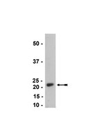
Recent progress in the structural identification of human melanoma antigens recognized by autologous cytotoxic T cells has led to the recognition of a new melanocyte differentiation antigen, Melan-A(MART-1). To determine the properties of the Melan-A gene product, Melan-A recombinant protein was produced in Escherichia coli and used to generate mouse monoclonal antibodies (mAbs). Two prototype mAbs, A103 and A355, were selected for detailed study. Immunoblotting results with A103 showed a 20-22-kDa doublet In Melan-A mRNA positive melanoma cell lines and no reactivity with Melan-A mRNA-negative cell lines. A355, in addition to the 20-22-kDa doublet, recognized several other protein species in Melan-A mRNA-positive cell lines. Immunocytochemical assays on cultured melanoma cells showed specific and uniform cytoplasmic staining in Melan-A mRNA-positive cell lines. Immunohistochemical analysis of normal human tissues with both mAbs showed staining of adult melanocytes and no reactivity with the other normal tissues tested. Analysis of 21 melanoma specimens showed homogenous staining of tumor cell cytoplasm in 16 of 17 Melan-A mRNA-positive cases and no reactivity with the three Melan-A mRNA-negative cases. | 8650193
 |


