Millipore Sigma Vibrant Logo

MABC949 Anti-Glycoprotein 78 Antibody, clone 3F3A

View Products on Sigmaaldrich.com
MABC949
200 µL  
Recuperando precio...
No pudo obtenerse el precio
La cantidad mínima tiene que ser múltiplo de
Maximum Quantity is
Al finalizar el pedido Más información
Ahorró ()
 
Solicitar precio
Disponibilidad a confirmar
Ingrese cantidad 
Suspendido
Cantidades limitadas disponibles
Debe confirmarse disponibilidad
    El resto: se avisará
      El resto: se avisará
      Se avisará
      Póngase en contacto con el Servicio de Atención al Cliente
      Contact Customer Service

      Ofertas especiales


      Póngase en contacto con el Servicio de Atención al Cliente

      Descripción

      Replacement Information

      Ofertas especiales

      Tabla espec. clave

      Species ReactivityKey ApplicationsHostFormatAntibody Type
      H, MWB, Function Assay, ICC, IHCRPurifiedMonoclonal Antibody
      Description
      Catalogue NumberMABC949
      DescriptionAnti-Glycoprotein 78 Antibody, clone 3F3A
      Alternate Names
      • E3 ubiquitin-protein ligase AMFR
      • AMF receptor
      • Autocrine motility factor receptor
      • gp78
      • RING finger protein 45
      Background InformationE3 ubiquitin-protein ligase AMFR (UniProt Q9UKV5; also known as AMF receptor, Autocrine motility factor receptor, gp78, RING finger protein 45) is encoded by the RNF45 (also known as AMFR) gene (Gene ID 267) in human. Glycoprotein 78 (gp78) is an endoplasmic reticulum (ER) membrane-anchored E3 ubiquitin ligase that plays a key role in ER-associated degradation (ERAD). Gp78 is localized to a mitochondria-associated ER domain, where it induces mitochondrial fragmentation by targeting the mitochondrial fusion proteins mitofusin 1 and 2 (Mfn1 and Mfn2, respectively) for degradation upon mitochondrial depolarization. Other known ERAD substrates processed by gp78 include ApoB lipoprotein, HMG CoA reductase, CD3-delta, cystic fibrosis transmembrane conductance regulator, the metastasis suppressor KAI1, unglycosylated prion protein (PrP), and the T-cell receptor. Gp78 is a 7-transmembrane protein (a.a. 82-102, 122-142, 145-165, 186-206, 215-235, 276-296, 429-449) with a RING-type Zinc finger (a.a. 341-379), a CUE domain (a.a. 456-498), and a VCP/p97-interacting motif (VIM; a.a. 622-640).
      References
      Product Information
      FormatPurified
      PresentationPurified rat IgM in buffer containing PBS without azide.
      Quality LevelMQ100
      Applications
      Application
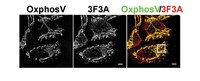
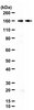
Detect GP78 using this rat monoclonal Anti-Glycoprotein 78, clone 3F3A, Cat. No. MABC949, validated for use in Function Assays, Immunocytochemistry, Immunohistochemistry, and Western Blotting.
      Key Applications
      • Western Blotting
      • Function Assay
      • Immunocytochemistry
      • Immunohistochemistry
      Application NotesImmunocytochemistry: A representative lot detected glycoprotein 78 immunoreactivity colocalized with that of the mitochondria marker OxPhosV in COS-7 cells (Courtesy of I. Robert Nabi, Ph.D., University of British Columbia, Canada).

      Affects Function: A representative lot stimulated the mobility of B16-F1 mouse melanoma cells on colloidal gold-coated surface (Nabi, I.R., et al. (1990). Cancer Res. 50(2):409-414).

      Immunocytochemistry Analysis: A representative lot detected both surface and cytoplasmic gp78 immunoreactivity by fluorescent immunocytochemistry staining of intact or MeOH/3% paraformaldehyde-fixed and 0.5% Triton X-100-permeablized mouse A31 fibroblasts and A31M angiosarcoma cells (Niinaka, Y., et al. (1998). Cancer Res. 58(12):2667-2674).

      Immunocytochemistry Analysis: Clone 3F3A ascites fluid immunolocalized gp78 on the surface of adherent B16-F1 mouse melanoma cells by fluorescent immunocytochemistry staining of 3% paraformaldehyde-fixed cells (Nabi, I.R., et al. (1990). Cancer Res. 50(2):409-414).

      Immunohistochemistry Analysis: A representative lot detected an elevated gp78 immunoreactivity in frozen mammary gland sections from 235RLF transgenic mice than age-matched wild type mice (Joshi, B., et al. (2010). J. Biol. Chem. 285(12):8830-8839).

      Western Blotting Analysis: A representative lot detected shRNA-mediated gp78 downregulation in HT-1180 human fibrosarcoma cells (Fu, M., et al. (2013). Mol. Biol. Cell. 24(8):1153-1162).

      Western Blotting Analysis: A representative lot detected endogenous gp78 and gp78 transgene expression in mammary glands from wild-type and transgenic mice. Clone 3F3A detected a higher gp78 expression in metastatic MDA-435 cells than in non-metastatic MCF7 breast carcinoma cells (Joshi, B., et al. (2010). J. Biol. Chem. 285(12):8830-8839).

      Western Blotting Analysis: A representative lot detected much higher gp78 expression in angiosarcoma (human HT-1180 and mouse A31M) than in fibroblast (human IMR90 and mouse A31) lines (Niinaka, Y., et al. (1998). Cancer Res. 58(12):2667-2674).

      Western Blotting Analysis: A representative lot detected gp78 and a ~150 kDa band in B16-F1 mouse melanoma cell extract. Autocrine motility factor (AMF) competed against clone 3F3A for binding the gp78 target band. Cell extract sialidase treatment decreased the antibody's immunoreactivity toward gp78 and abolished ~150 kDa band detection (Nabi, I.R., et al. (1990). Cancer Res. 50(2):409-414).
      Biological Information
      ImmunogenPeanut lectin (PNA) resin-purified membrane glycoprotein 78 from B16-F1 cells (Nabi, I.R., and Raz, A. (1987). Int. J. Cancer. 40(3):396-402).
      Epitopeextracellular domain
      Clone3F3A
      ConcentrationPlease refer to lot specific datasheet.
      HostRat
      SpecificityClone 3F3A detected glycoprotein 78 (gp78) and a ~150 kDa glycoprotein in B16-F1 mouse melanoma cell extract. Cell extract sialidase treatment decreased the antibody's immunoreactivity toward gp78 and abolished ~150 kDa band detection (Nabi, I.R., et al. (1990). Cancer Res. 50(2):409-414).
      IsotypeIgMκ
      Species Reactivity
      • Human
      • Mouse
      Antibody TypeMonoclonal Antibody
      Entrez Gene Number
      Gene Symbol
      • RNF45
      • AMFR
      UniProt Number
      Molecular Weight~78 kDa observed. Target band size appears larger than the calculated molecular weight of 73.00 kDa due to glycosylation. Uncharacterized bands may be observed in some lysate(s).
      Physicochemical Information
      Dimensions
      Materials Information
      Toxicological Information
      Safety Information according to GHS
      Safety Information
      Product Usage Statements
      Quality AssuranceWestern Blotting Analysis: A 1:62.5 dilution of this antibody detected glycoprotein 78 (gp78) in 50 µg of HeLa cell lysate.
      Usage Statement
      • Unless otherwise stated in our catalog or other company documentation accompanying the product(s), our products are intended for research use only and are not to be used for any other purpose, which includes but is not limited to, unauthorized commercial uses, in vitro diagnostic uses, ex vivo or in vivo therapeutic uses or any type of consumption or application to humans or animals.
      Storage and Shipping Information
      Storage ConditionsStable for 1 year at -20°C from date of receipt.
      Handling Recommendations: Upon receipt and prior to removing the cap, centrifuge the vial and gently mix the solution. Aliquot into microcentrifuge tubes and store at -20°C. Avoid repeated freeze/thaw cycles, which may damage IgG and affect product performance.
      Packaging Information
      Material Size200 µL
      Transport Information
      Supplemental Information
      Specifications
      Global Trade Item Number
      Número de referencia GTIN
      MABC949 04054839057151